. 1996 Dec 10;93(25):14474-9.
doi: 10.1073/pnas.93.25.14474.

The change in hydrogen bond strength accompanying charge rearrangement: implications for enzymatic catalysis

Affiliations

The change in hydrogen bond strength accompanying charge rearrangement: implications for enzymatic catalysis

S O Shan et al. Proc Natl Acad Sci U S A. .

Abstract

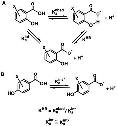
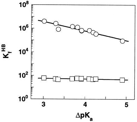
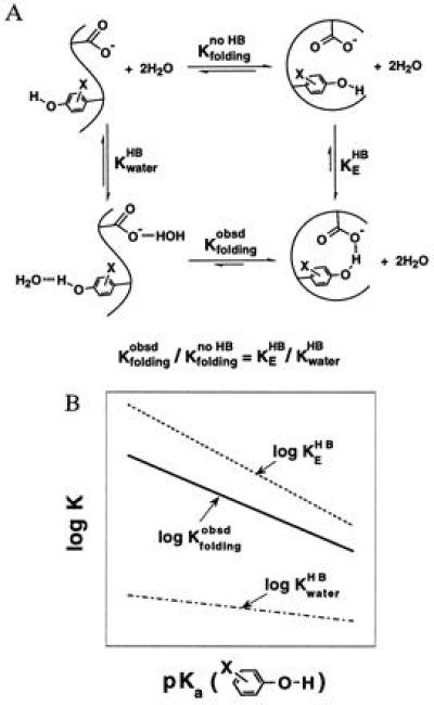
The equilibrium for formation of the intramolecular hydrogen bond (KHB) in a series of substituted salicylate monoanions was investigated as a function of delta pKa, the difference between the pKa values of the hydrogen bond donor and acceptor, in both water and dimethyl sulfoxide. The dependence of log KHB upon delta pKa is linear in both solvents, but is steeper in dimethyl sulfoxide (slope = 0.73) than in water (slope = 0.05). Thus, hydrogen bond strength can undergo substantially larger increases in nonaqueous media than aqueous solutions as the charge density on the donor or acceptor atom increases. These results support a general mechanism for enzymatic catalysis, in which hydrogen bonding to a substrate is strengthened as charge rearranges in going from the ground state to the transition state; the strengthening of the hydrogen bond would be greater in a nonaqueous enzymatic active site than in water, thus providing a rate enhancement for an enzymatic reaction relative to the solution reaction. We suggest that binding energy of an enzyme is used to fix the substrate in the low-dielectric active site, where the strengthening of the hydrogen bond in the course of a reaction is increased.


Figures

Scheme I
Scheme I
Scheme II
Scheme II
Figure 1
Figure 1
The dependence of log KHB upon ΔpKawater in DMSO (○) and H2O (□) for the H bond in substituted SA monoanions. The Brønsted slopes are 0.05 and 0.73 in water and DMSO, respectively. A common ΔpKa scale in water was used to allow a direct comparison of the magnitude of changes in H bond strength in the two solvents. Data are from Table 1.
Figure 2
Figure 2
A greater increase in H bond strength accompanying changes in the charge density of donor/acceptors in DMSO than in water. The logarithm of equilibrium constants for H bonding in DMSO are plotted against those in water for substituted salicylates (note the difference in scales). The solid line represents a least squares fit of the data and gives a slope of 15. The dashed line with a slope of unity, which represents the correlation expected if the strengthening of the H bond were the same in the two media, is shown for comparison. Data are from Table 1.
Figure 3
Figure 3
Diagrammatic illustration of potential catalysis that can be obtained from a greater strengthening of H bonds accompanying charge rearrangements at a nonaqueous enzymatic active site than in water, using the example of the TIM reaction. The pKa of the substrate carbonyl oxygen increases during the course of reaction, so that ΔpKa between this oxygen atom and the enzymatic or solution H bond donor (His-E or HOH, respectively) decreases. This leads to an increase in H bond strength. This increase would be greater at the enzymatic active site (ΔΔGE) than in solution (ΔΔGsoln) because of the greater Brønsted slopes in nonaqueous environments. The amount of catalysis obtained from this strategy, relative to the solution reaction, is ΔΔG = ΔΔGE − ΔΔGsoln.
Figure 4
Figure 4
A large Brønsted slope for H bonding in proteins is inferred from folding studies of Staphylococcal nuclease mutants. (A) Thermodynamic analysis depicting the effect of changing the strength of the H bond donor, the substituted tyrosine hydroxyl, on the stability of the protein. The folding equilibrium of a hypothetical non-H-bonded species (Kfoldingno HB) is used to dissect the effects from H bonding. (B) Schematic depiction of the dependences of H bonding and folding equilibria on the pKa value of substituted tyrosines. As the tyrosine hydroxyl becomes more acidic, the strengthening of its H bond to the enzymatic glutamate and to water stabilizes the folded and unfolded protein, respectively. The slope of the plot of log Kfoldingobsd versus pKa is the difference between the slopes of plots of log KEHB versus pKa and KwaterHB versus pKa. This follows from the thermodynamic relationship shown in A, which gives Δlog Kfoldingobsd = Δlog KEHB − Δlog KwaterHB (i.e., the greater strengthening of the H bond on the protein results in a change in the observed stability of the protein. Note that Δlog Kfoldingno HB = 0 by definition because the folding equilibrium between the non-H-bonded species does not depend on the strength of the H bond donor.

References

    1. Kresge A J. Pure Appl Chem. 1991;63:213–221.
    1. Bordwell F G. Acc Chem Res. 1988;21:456–463.
    1. Bordwell F G, Branca J C, Hughes D L, Olmstead W N. J Org Chem. 1980;45:3305–3313.
    1. Bordwell F G, McCallum R J, Olmstead W N. J Org Chem. 1984;49:1424–1427.
    1. Kortum G, Vogel W, Andrussow K. Dissociation Constants of Organic Acids in Aqueous Solution. London: Butterworths; 1961. pp. 352–464.

Publication types

LinkOut - more resources