. 1996 Dec 10;93(25):14526-31.
doi: 10.1073/pnas.93.25.14526.

Ordered water molecules as key allosteric mediators in a cooperative dimeric hemoglobin

Affiliations

Ordered water molecules as key allosteric mediators in a cooperative dimeric hemoglobin

W E Royer Jr et al. Proc Natl Acad Sci U S A. .

Abstract

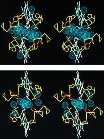
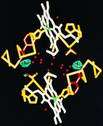
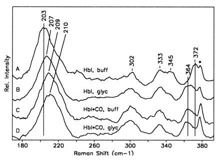
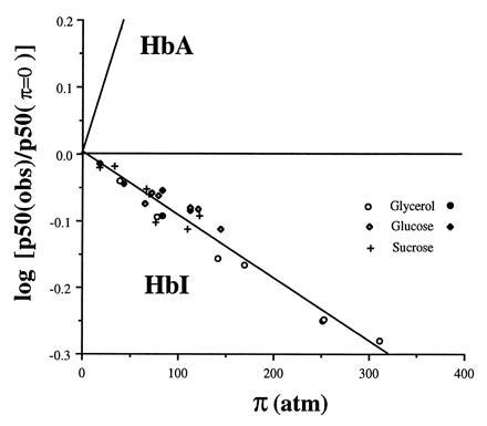
One of the most remarkable structural aspects of Scapharca dimeric hemoglobin is the disruption of a very well-ordered water cluster at the subunit interface upon ligand binding. We have explored the role of these crystallographically observed water molecules by site-directed mutagenesis and osmotic stress techniques. The isosteric mutation of Thr-72-->Val in the interface increases oxygen affinity more than 40-fold with a surprising enhancement of cooperativity. The only significant structural effect of this mutation is to destabilize two ordered water molecules in the deoxy interface. Wild-type Scapharca hemoglobin is strongly sensitive to osmotic conditions. Upon addition of glycerol, striking changes in Raman spectrum of the deoxy form are observed that indicate a transition toward the liganded form. Increased osmotic pressure, which lowers the oxygen affinity in human hemoglobin, raises the oxygen affinity of Scapharca hemoglobin regardless of whether the solute is glycerol, glucose, or sucrose. Analysis of these results provides an estimate of six water molecules lost upon oxygen binding to the dimer, in good agreement with eight predicted from crystal structures. These experiments suggest that the observed cluster of interfacial water molecules plays a crucial role in communication between subunits.


Figures

Figure 1
Figure 1
Stereo diagram depicting the interface water molecules of HbI viewed down the molecular dyad. Water molecules are shown as blue spheres representing the approximate van der Waals radii of oxygen atoms in (i) deoxy HbI (7) (Protein Data Bank code 4SDH) and (ii) oxy HbI (8) (Protein Data Bank code 1HBI). In addition to the water molecules, for both subunits an α carbon trace for residues 93–101 and 69–75 is shown in yellow, the heme groups are shown in white (with the heme iron green), side chains for the distal (residue 69) and proximal (residue 101) histidines are shown in yellow, while the side chains for Phe-97 are shown in red and the side chains for Thr-72 are shown in yellow with the hydroxyl shown in red. The deoxy water molecules are more well ordered than those of the oxy structure: the 19 deoxy water molecules shown have refined B factors that average 21.2 Å2 and average occupancy of 0.98, while the 11 shown for the oxy structure have an average B-factor of 24.3 Å2 and occupancy of 0.91. The deoxy interface water molecules form a clear cluster with five of the water molecules in position to form hydrogen bonds with other water molecules but not with protein atoms. In the oxy interface, only one water molecule has all its possible hydrogen bonds with other water molecules.
Figure 2
Figure 2
Difference Fourier map comparing wild-type HbI and T72V deoxy crystals. Atomic structure is shown as in Fig. 1, but with water molecules in red. Contours are drawn at 6σ for a map calculated between T72V and wild-type deoxy data (Protein Data Bank code R4SDHSF) using 10,578 reflections corresponding to spacings between 10- and 2.0-Å resolution. This indicates the only significant differences between the structures is the loss, or disordering, in T72V of the two water molecules normally in a position to hydrogen bond to the hydroxyl group of Thr-72 in each subunit.
Figure 3
Figure 3
Plot showing the low frequency resonance Raman spectra of deoxy HbI in aqueous buffer (pH 7.1, 100 mM phosphate) (line A); deoxy HbI in a glycerol/water solution (90% glycerol by volume) (line B); HbI*, the 10-ns photoproduct of HbICO in aqueous buffer (line C); and HbI* in a 90% glycerol/water solution (line D). The low frequency band occurring between 203 and 210 cm−1 is the iron-proximal histidine stretching mode. The bands occurring between 330 and 372 cm−1 are all sensitive to the propionate stretching motions. The shift in the propionate bands in going from deoxy HbI to HbI* in buffer is ascribed in this case to the large shift in the heme upon ligation (E.S.P., W.E.R., and J.M.F., unpublished data). It can be seen that the addition of glycerol to the deoxy sample produces a population that has almost the same iron-proximal histidine stretching and propionate frequencies as HbI*. The spectral feature marked by ∗ is from the sapphire window.
Figure 4
Figure 4
Plot showing the dependence of oxygen affinity with osmotic pressure for HbI and human hemoglobin (HbA). The value of p50 at π = 0 was estimated from measurements in buffer alone. The line showing dependence for human hemoglobin is obtained from Colombo et al. (25). Data points are shown for tonometer measurements with glycerol (○), glucose (⋄), and sucrose (+) solutions and Hem-O-Scan measurements for glycerol (•) and glucose (♦). Note the opposite direction for HbI, in which osmotic pressure increases oxygen affinity, compared with HbA, for which osmotic pressure decreases oxygen affinity. The slope of the least squares fit of the HbI data is −9.5 × 10⁻⁴ ± 0.5 atm−1, which indicates that an additional 6.2 ± 0.3 water molecules are bound to the deoxy state relative to the oxy state.

Similar articles

Cited by

References

    1. Karplus P A, Faerman C. Curr Opin Struct Biol. 1994;4:770–776.
    1. Badger J, Caspar D L D. Proc Natl Acad Sci USA. 1991;88:622–626. - PMC - PubMed
    1. Quiocho F A, Wilson D K, Vyas N K. Nature (London) 1989;340:404–407. - PubMed
    1. Singer P T, Smalås A, Carty R P, Mangel W F. Science. 1993;259:669–673. - PubMed
    1. Bhat T N, Bentley B A, Boulot G, Breene M I, Tello D, Dall’Acqua W, Souchon H, Schwarz F P, Mariuzza R A, Poljak R J. Proc Natl Acad Sci USA. 1994;91:1089–1093. - PMC - PubMed

Publication types

LinkOut - more resources