Cultured melanocytes from dilute mutant mice exhibit dendritic morphology and altered melanosome distribution
- PMID: 8962090
- PMCID: PMC26171
- DOI: 10.1073/pnas.93.25.14554
Cultured melanocytes from dilute mutant mice exhibit dendritic morphology and altered melanosome distribution
Abstract
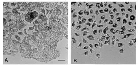
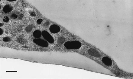
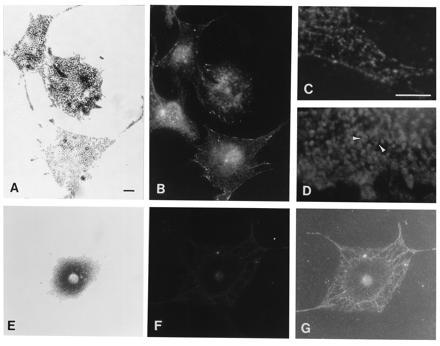
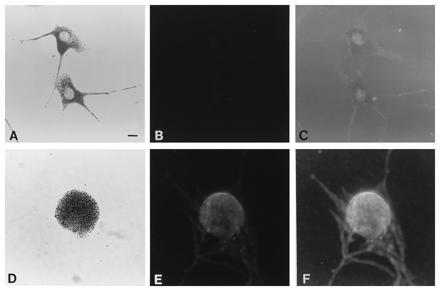
Mutant alleles at the dilute unconventional myosin heavy chain locus cause diluted coat color, opisthotonic seizures, and death. The dilute coat color phenotype is caused by irregular clumping of pigment in the hair, but amounts of melanin are unchanged from wild-type controls. The melanocyte phenotype has been described as adendritic, since hair bulb and Harderian gland melanocytes appear to be rounded in tissue sections. These observations do not exclude the possibility that the processes lack pigment, since the melanocyte shape was judged by the distribution of melanin. We have tested this hypothesis by culturing primary melanocytes from dilute mutant and wild-type mice. The mutant melanocytes do not lack processes; instead, they exhibit a concentrated perinuclear distribution of melanosomes, while wild-type melanocytes have a very uniform cytoplasmic distribution of melanosomes. Electron micrographs show no detectable differences in melanosome morphology or maturation between dilute and wild-type melanocytes. Immunofluorescence experiments indicate that the dilute protein is concentrated in regions of the cytoplasm that contain melanosomes. These experiments show that the dilute myosin is necessary for the localization of melanosomes, either by active transport or tethering.
Figures


Similar articles
-
Melanoregulin regulates a shedding mechanism that drives melanosome transfer from melanocytes to keratinocytes.Proc Natl Acad Sci U S A. 2012 Jul 31;109(31):E2101-9. doi: 10.1073/pnas.1209397109. Epub 2012 Jul 2. Proc Natl Acad Sci U S A. 2012. PMID: 22753477 Free PMC article.
-
The predominant defect in dilute melanocytes is in melanosome distribution and not cell shape, supporting a role for myosin V in melanosome transport.J Muscle Res Cell Motil. 1997 Oct;18(5):517-27. doi: 10.1023/a:1018659117569. J Muscle Res Cell Motil. 1997. PMID: 9350005
-
Visualization of melanosome dynamics within wild-type and dilute melanocytes suggests a paradigm for myosin V function In vivo.J Cell Biol. 1998 Dec 28;143(7):1899-918. doi: 10.1083/jcb.143.7.1899. J Cell Biol. 1998. PMID: 9864363 Free PMC article.
-
Making sense of melanosome dynamics in mouse melanocytes.Pigment Cell Res. 2000 Aug;13(4):241-7. doi: 10.1034/j.1600-0749.2000.130405.x. Pigment Cell Res. 2000. PMID: 10952391 Review.
-
The dilute locus and Griscelli syndrome: gateways towards a better understanding of melanosome transport.Pigment Cell Res. 2001 Oct;14(5):320-7. doi: 10.1034/j.1600-0749.2001.140503.x. Pigment Cell Res. 2001. PMID: 11601653 Review.
Cited by
-
Myosin Va movements in normal and dilute-lethal axons provide support for a dual filament motor complex.J Cell Biol. 1999 Sep 6;146(5):1045-60. doi: 10.1083/jcb.146.5.1045. J Cell Biol. 1999. PMID: 10477758 Free PMC article.
-
Role of Myosin Va in the plasticity of the vertebrate neuromuscular junction in vivo.PLoS One. 2008;3(12):e3871. doi: 10.1371/journal.pone.0003871. Epub 2008 Dec 5. PLoS One. 2008. PMID: 19057648 Free PMC article.
-
Mlc1p is a light chain for the unconventional myosin Myo2p in Saccharomyces cerevisiae.J Cell Biol. 1998 Aug 10;142(3):711-22. doi: 10.1083/jcb.142.3.711. J Cell Biol. 1998. PMID: 9700160 Free PMC article.
-
Molecular characterization of myosin V from Drosophila melanogaster.J Muscle Res Cell Motil. 1998 Feb;19(2):129-41. doi: 10.1023/a:1005356511634. J Muscle Res Cell Motil. 1998. PMID: 9536440
-
The role of the COOH terminus of Sec2p in the transport of post-Golgi vesicles.J Cell Biol. 2000 Apr 3;149(1):95-110. doi: 10.1083/jcb.149.1.95. J Cell Biol. 2000. PMID: 10747090 Free PMC article.
References
Publication types
MeSH terms
Substances
Associated data
- Actions
Grants and funding
LinkOut - more resources
Full Text Sources
Molecular Biology Databases