. 1996 Dec 10;93(25):14602-7.
doi: 10.1073/pnas.93.25.14602.

Mouse cytoplasmic polyadenylylation element binding protein: an evolutionarily conserved protein that interacts with the cytoplasmic polyadenylylation elements of c-mos mRNA

Affiliations

Mouse cytoplasmic polyadenylylation element binding protein: an evolutionarily conserved protein that interacts with the cytoplasmic polyadenylylation elements of c-mos mRNA

F Gebauer et al. Proc Natl Acad Sci U S A. .

Abstract

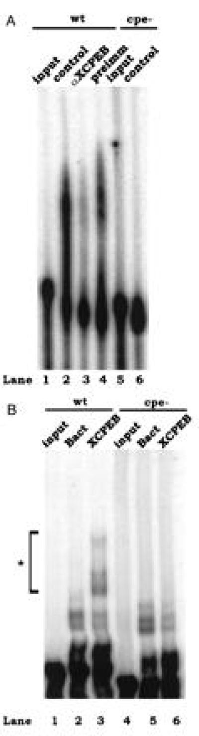
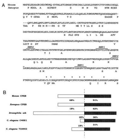
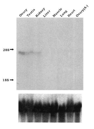
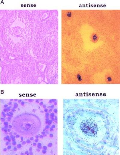
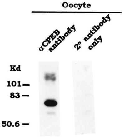
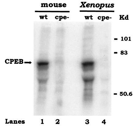
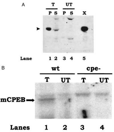
Cytoplasmic polyadenylylation is an essential process that controls the translation of maternal mRNAs during early development and depends on two cis elements in the 3' untranslated region: the polyadenylylation hexanucleotide AAUAAA and a U-rich cytoplasmic polyadenylylation element (CPE). In searching for factors that could mediate cytoplasmic polyadenylylation of mouse c-mos mRNA, which encodes a serine/threonine kinase necessary for oocyte maturation, we have isolated the mouse homolog of CPEB, a protein that binds to the CPEs of a number of mRNAs in Xenopus oocytes and is required for their polyadenylylation. Mouse CPEB (mCPEB) is a 62-kDa protein that binds to the CPEs of c-mos mRNA. mCPEB mRNA is present in the ovary, testis, and kidney; within the ovary, this RNA is restricted to oocytes. mCPEB shows 80% overall identity with its Xenopus counterpart, with a higher homology in the carboxyl-terminal portion, which contains two RNA recognition motifs and a cysteine/histidine repeat. Proteins from arthropods and nematodes are also similar to this region, suggesting an ancient and widely used mechanism to control polyadenylylation and translation.


Figures

Figure 1
Figure 1
Xenopus CPEB recognizes the mouse c-mos CPEs. (A) Polyadenylylation of c-mos mRNA 3′-UTR in Xenopus egg extracts. Radiolabeled c-mos 3′-UTR was incubated with egg extracts that were untreated (control), mock-depleted (preimm), or immunodepleted of CPEB (anti-XCPEB, αXCPEB). The RNA was then extracted and analyzed for polyadenylylation by denaturing gel electrophoresis and autoradiography. Polyadenylylation assays were performed with wild-type RNA (lanes 1–4) or a mutant RNA that lacked CPEs (lanes 5 and 6). (B) Gel mobility-shift assay with His-tagged XCPEB. Radiolabeled wild-type (lanes 1–3) or cpe− (lanes 4–6) c-mos 3′-UTR was incubated with protein preparations from bacteria expressing (lanes 3 and 6) or lacking (lanes 2 and 5) XCPEB or was incubated without protein (lanes 1 and 4). The RNA was resolved in a 4% polyacrylamide gel containing 1 M urea.
Figure 2
Figure 2
mCPEB amino acid sequence and its comparison with related proteins. (A) Predicted amino acid sequence of mouse CPEB compared with Xenopus CPEB. The conserved ribonucleoprotein-type RNA recognition motifs are denoted. The asterisks indicate the cysteine and histidine residues that could form a zinc finger. (B) Schematic representation of a comparison between mCPEB and related proteins from other organisms. The numbers indicate the percentage of amino acid identity of the various proteins with respect to mCPEB. The highest identity (solid box) is found in the carboxyl-terminal portion.
Figure 3
Figure 3
Tissue distribution of mCPEB mRNA. (Upper) Fifty micrograms of total RNA from the indicated mouse tissues was examined by Northern blot analysis using a random-primed probe complementary to nt 898-2610 of mCPEB cDNA. Ovary RNA that was depleted of poly(A)+ RNA [ovary (A−)] was included as a negative control. The positions of the 18S and 28S ribosomal RNAs are indicated as markers at the left. To ensure that equivalent amounts of RNA were loaded in each lane, the filter was stripped and rehybridized with a probe complementary to the 18S ribosomal RNA (Lower).
Figure 4
Figure 4
Distribution of mCPEB mRNA in the ovary. Low (A) and high (B) magnifications of an in situ hybridization to ovary sections, using as a probe an 35S-labeled RNA complementary to nt 1424–1520 of mCPEB cDNA (antisense). The sense labeled RNA was used as a negative control.
Figure 5
Figure 5
mCPEB is present in oocytes. Immunoblot of a mouse oocyte extract with affinity-purified anti-XCPEB antibody. An extract derived from 385 oocytes was used. As a negative control, a similar extract was only incubated with the secondary antibody. The protein bands were visualized by enhanced chemiluminescence.
Figure 6
Figure 6
Endogenous mCPEB recognizes the CPEs of c-mos mRNA. Radiolabeled c-mos mRNA 3′-UTR containing (wt) or lacking (cpe−) CPEs was incubated with protein extracts from 265 mouse oocytes (lanes 1 and 2) or one Xenopus oocyte (lanes 3 and 4). After an incubation of 30 min at room temperature, the extract was UV-irradiated, digested with RNase A, and immunoselected with anti-XCPEB antibody. Radiolabeled proteins were resolved by SDS/polyacrylamide gel electrophoresis and visualized using a PhosphorImager.
Figure 7
Figure 7
Recombinant mCPEB recognizes the CPEs of mouse c-mos mRNA. (A) Immunoblot of mCPEB expressed by transient transfection of Cos cells. Cells were transfected with mCPEB cDNA (T) or mock-transfected (UT, no DNA). After an incubation of 36 h, the cells were homogenized and the nuclei were pelleted by centrifugation. Equivalent amounts of pellet (P) and supernatant (S) (approximately one-third of a 100-mm diameter dish) were loaded in a 10% polyacrylamide gel and examined by Western blot analysis using affinity-purified anti-XCPEB antibody. A protein extract corresponding to one-fourth of a Xenopus oocyte (X) was used as a positive control. The size of CPEB is indicated by an arrowhead at the left. (B) UV-crosslinking and immunoselection of mCPEB expressed in Cos cells. Protein extracts from Cos cells that were transfected with mCPEB cDNA (T) or mock-transfected (UT) were UV-crosslinked to radiolabeled c-mos 3′-UTR containing (wt) or lacking (cpe−) CPEs. A supernatant corresponding to 50% of a 100-mm dish was used per lane. Crosslinked products were immunoselected with anti-XCPEB antibody and resolved by SDS/polyacrylamide gel electrophoresis. The results were visualized in a PhosphorImager.

References

    1. Richter J. In: Translational Control. Hershey J W B, Mathews M B, Sonenberg N, editors. Plainview, NY: Cold Spring Harbor Lab. Press; 1996. pp. 481–503.
    1. Vassalli J, Stutz A. Curr Biol. 1995;5:476–479. - PubMed
    1. Sallés F, Lieberfarb M, Wreden C, Gergen J, Strickland S. Science. 1994;266:1996–1998. - PubMed
    1. Simon R, Wu L, Richter J D. Dev Biol. 1996;179:239–250. - PubMed
    1. Gebauer F, Xu W, Cooper G, Richter J. EMBO J. 1994;13:5712–5720. - PMC - PubMed

Publication types

Associated data