. 1996 Dec 10;93(25):14714-9.
doi: 10.1073/pnas.93.25.14714.

Ocular cicatricial pemphigoid antigen: partial sequence and biochemical characterization

Affiliations

Ocular cicatricial pemphigoid antigen: partial sequence and biochemical characterization

S Tyagi et al. Proc Natl Acad Sci U S A. .

Abstract

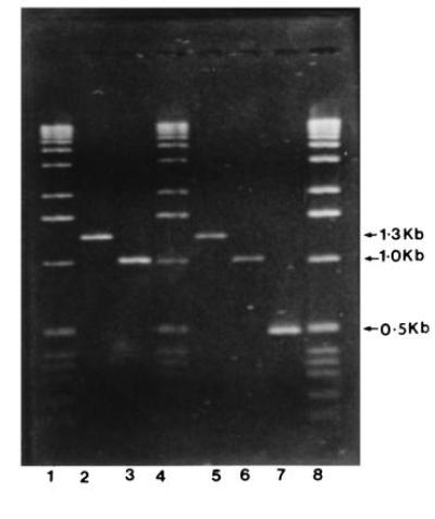
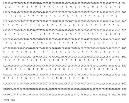
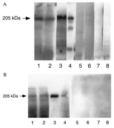
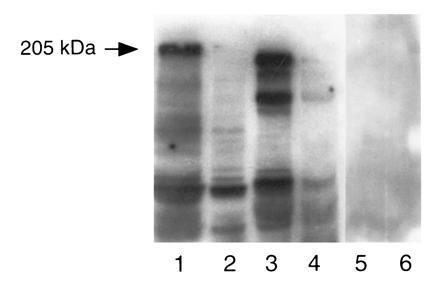
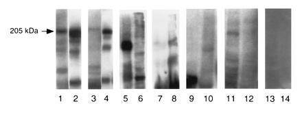
Ocular cicatricial pemphigoid (OCP) is an autoimmune disease that affects mainly conjunctiva and other squamous epithelia. OCP is histologically characterized by a separation of the epithelium from underlying tissues within the basement membrane zone. Immunopathological studies demonstrate the deposition of anti-basement membrane zone autoantibodies in vivo. Purified IgG from sera of patients with active OCP identified a cDNA clone from a human keratinocyte cDNA library that had complete homology with the cytoplasmic domain of beta 4-integrin. The sera recognized a 205-kDa protein in human epidermal, human conjunctiva, and tumor cell lysates that was identified as beta 4-integrin by its reaction with polyclonal and monoclonal antibodies to human beta 4-integrin. Sera from patients with bullous pemphigoid, pemphigus vulgaris, and cicatricial pemphigoid-like diseases did not recognize the 205-kDa protein, indicating the specificity of the binding. These data strongly implicate a role for human beta 4-integrin in the pathogenesis of OCP. It should be emphasized that multiple antigens in the basement membrane zone of squamous epithelia may serve as targets for a wide spectrum of autoantibodies observed in vesiculobullous diseases. Molecular definition of these autoantigens will facilitate the classification and characterization of subsets of cicatricial pemphigoid and help distinguishing them from bullous pemphigoid. This study highlights the function and importance of beta 4-integrin in maintaining the attachment of epithelial cells to the basement membrane.


Figures

Figure 1
Figure 1
PCR amplification of clone 10 of OCP antigen. Lanes: 1, 4, 0.5 μg of DNA ladder (GIBCO/BRL); 8, 1.0 μg of DNA ladder; 2 and 5, clone 8 (1.3 kb) as internal standard; 3 and 6, replicates of clone 10 (1.0 kb, OCP antigen clone); 7, internal standard for PCR amplification kit (Perkin–Elmer and CLONTECH).
Figure 2
Figure 2
Partial nucleotide sequence and deduced amino acid sequence of clone 10 of OCP antigen. Nucleotide sequence was analyzed on an Applied Biosystems model 373A automated DNA sequencer and deduced amino acid were performed at National Center for Biotechnology Information using blast network service. Nucleotide sequences from positions 42 to 964 have complete homology with published sequence of human β4-integrin.
Figure 3
Figure 3
(A) Immunoblot of OCP antigen/β4-integrin (205/210 kDa) using human epidermal, human conjunctival, and tumor cell lines (MDA 435 and UM-SCC-22) and detected by polyclonal antibody to β4-integrin. Lanes: 1, HEL; 2, HCL; 3, MDA 435 cell lysates; 4, UM-SCC-22 cell lysate reacted with polyclonal antibody to β4-integrin; 5–8, same lysates reacted with normal human serum. In all four groups (lanes 1–4), a 205/210-kDa protein band was observed when β4-integrin antibody was used. No similar protein band was detected when normal human serum was used (lanes 5–8). (B) The identical setup of experiment was used except lanes 3 and 4 were UM-SCC-22 and MDA 435, respectively, and monoclonal anti-β4-integrin was used instead of polyclonal.
Figure 4
Figure 4
Preabsorption/blocking studies by OCP patients’ sera using MDA 435 and UM-SCC-22 cell lysates. MDA 435 and UM-SCC-22 cell were preabsorbed with OCP patients’ sera and normal human serum that was previously bound to cyanogen bromide-activated Sepharose 4B. Samples were prepared for SDS/PAGE, transferred to nitrocellulose, examined by immunoblot analysis with β4-integrin polyclonal antibody and normal human serum. Lanes 1 and 2 contain MDA 435 cell lysate. In lane 1 lysate was absorbed with normal human serum (NHS) and then blotted with anti-β4-antibody. Note the 205-kDa band. In lane 2, cell lysate was first absorbed with OCP sera then blotted with anti-β4 antibody. Note the absence of the 205-kDa band. Lanes 3 and 4 represent UM-SCC-22 cell lysate treated similarly. Lanes 5 and 6 tumor cell lysates preabsorbed with NHS then reacted with NHS.
Figure 5
Figure 5
Specificity experiment using MDA 435 and UM-SCC-22 cell lysates and detected by various patients sera and anti-β4-integrin antibody. Lanes: 1, β4-integrin antibody; 3, OCP patients’ sera; 5, polyclonal anti-laminin 5; 7, anti-epiligrin human sera; 9, PV patients’ sera; 11, BP patients’ sera; 13, normal human sera. All these sera reacted with MDA cell lysates. Lanes: 2, β4-integrin antibody; 4, OCP patients’ sera; 6, anti-laminin 5; 8, anti-epiligrin human sera; 10, PV patients’ sera; 12, BP patients’ sera; 14, normal human sera. All these sera reacted with UM-SCC-22 cell lysates.

References

    1. Foster C S. Trans Am Ophthalmol Soc. 1986;84:527–663. - PMC - PubMed
    1. Ahmed A R, Hombal S M. Int J Dermatol. 1986;25:90–96. - PubMed
    1. Bernard P, Prost C, Aucoutuier P, Durepaire N, Denis F, Jean-Marie B. J Invest Dermatol. 1991;97:259–263. - PubMed
    1. Neiboer C, Boarsma D M, Woederma M J. Br J Dermatol. 1982;106:419–422. - PubMed
    1. Shimuzu H, Masunago T, Ishiko A, Matsumura K, Hashimoto T, Nishokawa T, Damloge-Hultsch N, Lazarova Z, Yancey K B. J Invest Dermatol. 1995;104:370–373. - PubMed

Publication types