

Molecular cloning and characterization of a murine pre-B-cell growth-stimulating factor/stromal cell-derived factor 1 receptor, a murine homolog of the human immunodeficiency virus 1 entry coreceptor fusin
- PMID: 8962122
- PMCID: PMC26203
- DOI: 10.1073/pnas.93.25.14726
Molecular cloning and characterization of a murine pre-B-cell growth-stimulating factor/stromal cell-derived factor 1 receptor, a murine homolog of the human immunodeficiency virus 1 entry coreceptor fusin
Abstract
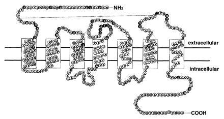
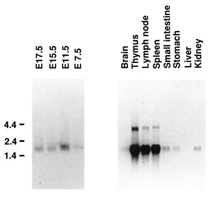
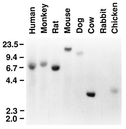
Pre-B-cell growth-stimulating factor/ stromal cell-derived factor 1 (PBSF/SDF-1) is a member of the CXC group of chemokines that is initially identified as a bone marrow stromal cell-derived factor and as a pre-B-cell stimulatory factor. Although most chemokines are thought to be inducible inflammatory mediators, PBSF/SDF-1 is essential for perinatal viability,. B lymphopoiesis, bone marrow myelopoiesis, and cardiac ventricular septal formation, and it has chemotactic activities on resting lymphocytes and monocytes. In this paper, we have isolated a cDNA that encodes a seven transmembrane-spanning-domain receptor, designated pre-B-cell-derived chemokine receptor (PB-CKR) from a murine pre-B-cell clone, DW34. The deduced amino acid sequence has 90% identity with that of a HUMSTSR/fusin, a human immunodeficiency virus 1 (HIV-1) entry coreceptor. However, the second extracellular region has lower identity (67%) compared with HUMSTSR/fusin. PB-CKR is expressed during embryo genesis and in many organs and T cells of adult mice. Murine PBSF/SDF-1 induced an increase in intracellular free Ca2+ in DW34 cells and PB-CKR-transfected Chinese hamster ovary (CHO) cells, suggesting that PB-CKR is a functional receptor for murine PBSF/SDF-1. Murine PBSF/ SDF-1 also induced Ca2+ influx in fusin-transfected CHO cells. On the other hand, considering previous results that HIV-1 does not enter murine T cells that expressed human CD4, PB-CKR may not support HIV-1 infection. Thus, PB-CKR will be an important tool for functional mapping of HIV-1 entry coreceptor fusin and for understanding the function of PBSF/SDF-1 further.
Figures

References
Publication types
MeSH terms
Substances
Associated data
- Actions
LinkOut - more resources
Full Text Sources
Other Literature Sources
Molecular Biology Databases
Research Materials
Miscellaneous
