. 1996 Dec 10;93(25):14736-41.
doi: 10.1073/pnas.93.25.14736.

Transfected Drosophila cells as a probe for defining the minimal requirements for stimulating unprimed CD8+ T cells

Affiliations

Transfected Drosophila cells as a probe for defining the minimal requirements for stimulating unprimed CD8+ T cells

Z Cai et al. Proc Natl Acad Sci U S A. .

Abstract

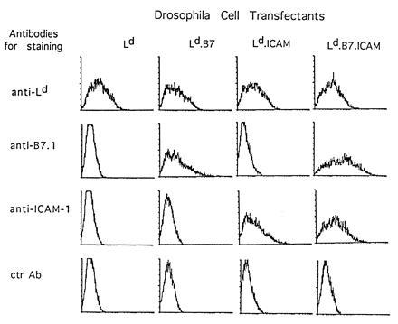
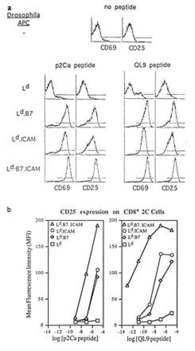
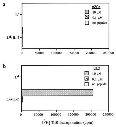
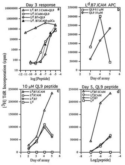
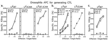
Stimulation of naive T cells by antigen-presenting cells (APC) is thought to involve two qualitatively different signals: signal one results from T-cell receptor (TCR) recognition of antigenic peptides bound to major histocompatibility complex (MHC) molecules, whereas signal two reflects contact with one or more costimulatory molecules. The requirements for stimulating naive T cells were studied with MHC class I-restricted CD8+ T cells from a T-cell receptor transgenic line, with defined peptides as antigen and transfected Drosophila cells as APC. Three main findings are reported. First, stimulation of naive T cells via signal one alone (MHC plus peptide) was essentially nonimmunogenic; thus T cells cultured with peptides presented by MHC class I-transfected Drosophila APC lacking costimulatory molecules showed little or no change in their surface phenotype. Second, cotransfection of two costimulatory molecules, B7-1 and intercellular adhesion molecule 1 (ICAM-1), converted class I+ Drosophila cells to potent APC capable of inducing strong T-proliferative responses and cytokine (interleukin 2) production. Third, B7-1 and ICAM-1 acted synergistically, indicating that signal two is complex; synergy between B7-1 and ICAM-1 varied from moderate to extreme and was influenced by both the dose and affinity of the peptide used and the parameter of T-cell activation studied. Transfected Drosophila cells are thus a useful tool for examining the minimal APC requirements for naive T cells.


Figures

Figure 1
Figure 1
Expression of Ld, B7-1, and ICAM-1 by transfected Drosophila cells. Drosophila cells were transfected with cDNAs encoding Ld, β2-microglobulin, B7-1, and ICAM-1 under the control of the metallothionein promoter as described. Transfected cells were separated with a FACS to obtain cells expressing Ld molecules and were maintained in vitro. The data show Ld, B7-1, and ICAM-1 expression on the cell lines 24 hr after induction with 1 mM CuSO4. The higher staining for B7-1 on Ld.B7.ICAM than Ld.B7 cells was not seen in other experiments.
Figure 2
Figure 2
Expression of activation markers on CD8+ 2C cells is influenced by peptide and costimulation. (a) Expression of CD69 and CD25 on CD8+ 2C cells stimulated with 10 μM peptides presented by transfected Drosophila cells. Purified CD8+ 2C cells were incubated with transfected Drosophila cells plus p2Ca or QL9 peptides (10 μM) in bulk (2 ml) culture for 12 hr and then stained for the markers shown. The data show staining of gated CD8+ cells. (Top) Staining of noncultured 2C cells is shown as a control. (b) Influence of peptide dose on expression of CD25 on CD8+ 2C cells. Purified CD8+ 2C cells were cultured with the indicated concentration of peptides presented by Drosophila cell APC for 12 hr and then stained for CD25 expression on gated CD8+ cells.
Figure 3
Figure 3
IL-2-dependent proliferative responses of CD8+ 2C cells to peptides presented by Drosophila cells transfected with Ld only. Responses to p2Ca (a) and QL9 (b) peptides were measured by culturing 5 × 10⁴ purified CD8+ 2C cells with 2 × 10⁵ Drosophila cells in the presence or absence of the indicated concentrations of peptides for 3 days. [3H]thymidine was added during the last 8 hr of culture; recombinant IL-2 was added at a final concentration of 20 units/ml. The data refer to the mean of triplicate cultures.
Figure 4
Figure 4
Features of proliferative responses of CD8+ 2C cells to peptides presented by transfected Drosophila cells. (a) Influence of peptide concentration on the day 3 response of CD8+ 2C cells to different Drosophila APC. (b) Influence of the dose of QL9 peptide on the kinetics of the response of CD8+ 2C cells with Ld.B7.ICAM APC. (c) Kinetics of the response of CD8+ 2C cells to 10 μM QL9 peptide presented by different Drosophila APC. (d) Influence of peptide dose on the day 5 response of CD8+ 2C cells with different Drosophila APC. CD8+ cells (5 × 10⁴) were cultured with 2 × 10⁵ Drosophila APC. The data show the mean of triplicate cultures.
Figure 5
Figure 5
Generation of primary CTL from naive CD8+ 2C cells cultured with QL9 peptide presented by Drosophila cell APC. (a) CTL activity of CD8+ 2C cells stimulated with Ld.B7, Ld.B7.ICAM, or Ld.ICAM APC plus QL9 peptide (10 μM) in the absence of exogenous cytokines. (b Left) CTL activity of CD8+ 2C cells stimulated with Ld.B7 APC plus QL9 peptide (10 μM) in the absence or presence of anti-IL-2 and/or anti-IL-4 mAb (2 μg/ml). (b Right) CTL activity of CD8+ 2C cells stimulated with Ld.ICAM APC plus QL9 peptide (10 μM) in the absence or presence of recombinant IL-2 (20 unit/ml). (c) CTL activity of CD8+ 2C cells stimulated with Ld.B7 APC plus QL9 peptide in the absence or presence of either anti-IL-2 or anti-IL-4 mAb (20 μg/ml). Purified CD8+ 2C cells (5 × 10⁵) were cultured with 2 × 10⁶ Drosophila APC. After 4 days, the cells were harvested and CTL activity was tested against [51Cr]-labeled RMA-S.Ld target cells loaded with p2Ca or control P1A.35-43 peptide (32); P1A peptide binds strongly to Ld but has no detectable affinity for the 2C TCR (32). The data show the mean level of specific 51Cr release from duplicate cultures.

References

    1. Germain R N. In: Fundamental Immunology. 3rd Ed. Paul W E, editor. New York: Raven; 1993. pp. 629–676.
    1. Janeway C A, Bottomly K. Cell. 1994;76:275–285. - PubMed
    1. Shimizu Y, van Seventer G A, Horgan K J, Shaw S. Immunol Rev. 1990;114:109–143. - PubMed
    1. Schwartz R H. Cell. 1992;71:1065–1068. - PubMed
    1. Linsley P S, Brady W, Grosmaire L, Aruffo A, Damle N K, Ledbetter J A. J Exp Med. 1991;173:721–730. - PMC - PubMed

Publication types

MeSH terms

Substances

LinkOut - more resources