. 1996 Dec 10;93(25):14821-6.
doi: 10.1073/pnas.93.25.14821.

Phenotype of arylsulfatase A-deficient mice: relationship to human metachromatic leukodystrophy

Affiliations

Phenotype of arylsulfatase A-deficient mice: relationship to human metachromatic leukodystrophy

B Hess et al. Proc Natl Acad Sci U S A. .

Abstract

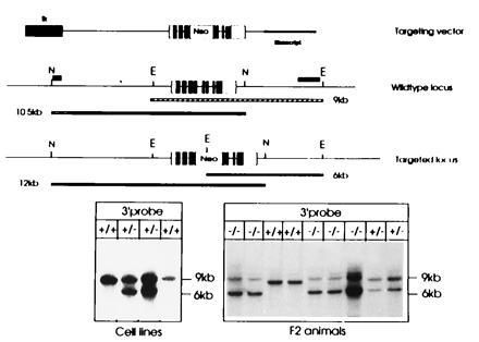
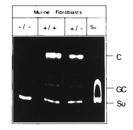
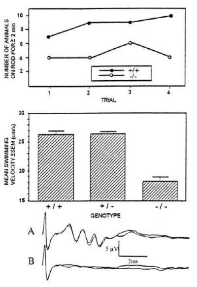
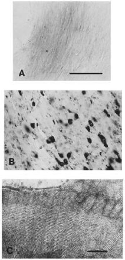
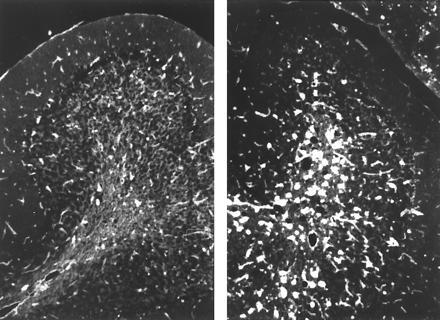
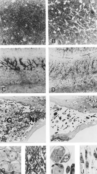
Metachromatic leukodystrophy is a lysosomal sphingolipid storage disorder caused by the deficiency of arylsulfatase A. The disease is characterized by progressive demyelination, causing various neurologic symptoms. Since no naturally occurring animal model of the disease is available, we have generated arylsulfatase A-deficient mice. Deficient animals store the sphingolipid cerebroside-3-sulfate in various neuronal and nonneuronal tissues. The storage pattern is comparable to that of affected humans, but gross defects of white matter were not observed up to the age of 2 years. A reduction of axonal cross-sectional area and an astrogliosis were observed in 1-year-old mice; activation of microglia started at 1 year and was generalized at 2 years. Purkinje cell dendrites show an altered morphology. In the acoustic ganglion numbers of neurons and myelinated fibers are severely decreased, which is accompanied by a loss of brainstem auditory-evoked potentials. Neurologic examination reveals significant impairment of neuromotor coordination.


Figures

Figure 1
Figure 1
Structure of the targeting vector and the targeted ASA locus. The structure of the linearized replacement vector is shown on top. The ASA gene is indicated by boxes, open parts are 5′ or 3′ untranslated regions, and solid parts depict coding sequences. The tk gene at the 5′ end is shown as a hatched box; the Bluescript plasmid vector is indicated by a bold line. The wild-type locus is shown below, N and E indicate NsiI and EcoRI sites, respectively. The position of external probes used to detect the targeted locus is shown at the 5′ and 3′ end. Size of the fragments (9 and 10.5 kb) hybridizing to these probes is indicated by the bars below. The third schematic drawing depicts the homologously recombined locus and the altered size of the DNA fragments detected by the 5′ and 3′ probe. Southern Blot analysis of DNA isolated from ES cells (Left) and tail tips of F2 animals (Right) are shown at the bottom. EcoRI-digested DNA was hybridized to the 3′ external probe. The size of fragments is indicated on the right. Genotypes are shown on top. This analysis was frequently complicated by the presence of an additional fragment slightly larger than the 9-kb fragment indicative of the wild-type locus. This fragment is most likely due to partial digestion as can be concluded from the varying intensities of this fragment in comparison to the 6-kb fragment in the homozygous −/− mice. Blots of NsiI-digested DNA hybridized with the 5′ probe are not shown.
Figure 2
Figure 2
Sulfatide loading of cultured fibroblasts. Cultured tail fibroblasts of −/−, +/−, and +/+ mice were exposed to fluorescently labeled sulfatide. Cells were allowed to metabolize the lipid for 16 h. Extracted lipids were subjected to thin layer chromatography and visualized by UV-irradiation. Lane Su contains only sulfatide. Su, nonmetabolized sulfatide; GC, galactocerebroside; C, ceramide.
Figure 3
Figure 3
Summary of the neuromotor examinations of control and deficient mice. The figure summarizes the results of tests in which neuromotor abnormalities were detected in the ASA-deficient mice. (Upper) Number of mice able to stay on a rotating rod for longer than 2 min. In each group, 10 mice were examined. Number of subsequent trials is indicated on the abscissa. (Middle) Swimming velocity of control and ASA-deficient mice. (Lower) Control (trace A) and deficient animals (trace B) were placed in front of a loudspeaker emitting clicks at a frequency of 11 Hz. Brainstem auditory-evoked potentials were recorded from electrodes placed in the anesthesized mice. Each curve represents 2000 measurements.
Figure 4
Figure 4
Histochemistry and ultrastructure of sulfatide storage. White matter (hippocampal fimbria) sections of a control (A) and ASA-deficient mouse (B) were stained for sulfatide with alcian blue. No staining can be detected in the brain of the control mouse. White matter of the deficient mouse displays cells filled with large storage granules and linearly arranged small cellular processes with small granules. (Bar = 100 μm.) (C) Ultrastructure of storage material. Prismatic herringbone-like inclusions in an astrocyte of a deficient mouse. (Bar = 100 nm.)
Figure 5
Figure 5
Activation of microglial cells in 1- and 2-year-old animals. Cerebellar sections of 1 (Left) and 2-year-old (Right) animals are shown. Sections show binding of RCA I, a marker of microglial activation.
Figure 6
Figure 6
Pathology of optic nerve, cerebellum, and acoustic ganglion. Transverse sections of the optic nerve of control (A) and ASA-deficient mouse (B) were stained with p-phenylenediamine. Myelin sheaths appear dark whereas axons and glial cells appear pale. Nonstained astrocytes appear as white patches, the number and size of which is clearly increased in the deficient animal. The cells feature thickened and elongated processes. Purkinje cells (C and D) were visualized by immunohistochemistry using an antibody to brain isoform of NO synthase. Cerebellar cortex of a control mouse is shown in C and that of a deficient mouse in D. Section of the inner ear of a control (E) and ASA-deficient (F) 11-month-old mouse. Ganglion cells (G) and myelinated nerve fibers (NF) in the acoustic ganglion are severely reduced in the deficient mice. Some lipid-laden macrophages (MP) are observed. Arrows depict surviving neurons. Details of perisomatic (G and I) and periaxonal (H and J) Schwann cells of control (G and H) and ASA (−/−) mice (I and J). In the deficient mouse, Schwann cells are swollen and filled with abnormal inclusions.

References

    1. Kolodny E H. In: The Metabolic and Molecular Bases of Inherited Disease. 6th Ed. Scriver C R, Beaudet A L, Sly W S, Valle D, editors. New York: McGraw–Hill; 1995. pp. 2693–2740.
    1. Wolfe H J, Pietra G G. Am J Pathol. 1964;44:921–930. - PMC - PubMed
    1. Gieselmann V, Zlotogora J, Harris A, Wenger D A, Morris C P. Hum Mutation. 1994;4:233–242. - PubMed
    1. Polten A, Fluharty A L, Fluharty C B, Kappler J, von Figura K, Gieselmann V. N Engl J Med. 1991;324:18–22. - PubMed
    1. Leinekugel P, Michel S, Conzelmann E, Sandhoff K. Hum Genet. 1992;88:513–523. - PubMed

Publication types

Substances