. 1996 Dec 10;93(25):14827-32.
doi: 10.1073/pnas.93.25.14827.

Guanylyl cyclase C is a selective marker for metastatic colorectal tumors in human extraintestinal tissues

Affiliations

Guanylyl cyclase C is a selective marker for metastatic colorectal tumors in human extraintestinal tissues

S L Carrithers et al. Proc Natl Acad Sci U S A. .

Abstract

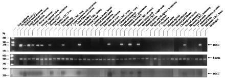
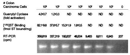
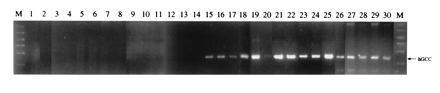
Guanylyl cyclase C (GCC) has been detected only in intestinal mucosa and colon carcinoma cells of placental mammals. However, this receptor has been identified in several tissues in marsupials, and its expression has been suggested in tissues other than intestine in placental mammals. Selective expression of GCC by colorectal tumor cells in extraintestinal tissues would permit this receptor to be employed as a selective marker for metastatic disease. Thus, expression of GCC was examined in human tissues and tumors, correlating receptor function with detection by PCR. GCC was detected by ligand binding and catalytic activation in normal intestine and primary and metastatic colorectal tumors, but not in extraintestinal tissues or tumors. Similarly, PCR yielded GCC-specific amplification products with specimens from normal intestine and primary and metastatic colorectal tumors, but not from extraintestinal tissues or tumors. Northern blot analysis employing GCC-specific probes revealed an approximately 4-kb transcript, corresponding to recombinant GCC, in normal intestine and primary and metastatic colorectal tumors, but not in extraintestinal tissues. Thus, GCC is selectively expressed in intestine and colorectal tumors in humans and appears to be a relatively specific marker for metastatic cancer cells in normal tissues. Indeed, PCR of GCC detected tumor cells in blood from some patients with Dukes B colorectal cancer and all patients examined with Dukes C and D colorectal cancer, but not in that from normal subjects or patients with Dukes A colon carcinoma or other nonmalignant intestinal pathologies.


Figures

Figure 1
Figure 1
Expression of GCC in tissues detected by RT-PCR. (Top and Middle) RT-PCR was performed employing mRNA purified from indicated tissues and primers for GCC (Top) or β-actin (control; Middle), as described. (Bottom) Aliquots of the above RT-PCRs were subjected to the ribonuclease protection assay as described. Left lane indicates size markers. Arrows indicate the size of human GCC (hGCC; ≈250 bp) and β-actin (≈600 bp) predicted from their defined sequences.
Figure 2
Figure 2
Sensitivity of enzyme activation, receptor binding, and RT-PCR to detect colon carcinoma cells in blood. The indicated number of T84 human colon carcinoma cells were diluted with 106 normal human blood cells in 1 ml and examined for ST (1 μM) activation of guanylyl cyclase and 125I-labeled ST (2.5 × 10⁻⁹ M) binding and by RT-PCR using GCC-specific nested primers, as described. PCR was conducted with 32P-labeled nucleotides, and the resultant amplification products resolved by electrophoresis were stained with ethidium bromide (lower row) or directly quantified (upper row). Fold activation was calculated as (cGMP produced with ST)/cGMP produced without ST). Values represent the mean ± SEM. ND, not detectable.
Figure 3
Figure 3
Detection of colon carcinoma cells in blood of patients using RT-PCR and GCC-specific nested primers. Total RNA was extracted from a 1-ml aliquot of blood cells, mRNA-purified, and used for RT-PCR, using GCC-specific nested primers, as described. Border lanes (M) indicate size markers (200–1000 bp). Arrow indicates the predicted size of the amplification product for human GCC (hGCC; ≈250 bp). Lane 1, normal volunteers (n = 10); lane 2, patients with a colonic adenomatous polyp (n = 6); lane 3, patient with an acute exacerbation of inflammatory bowel disease (n = 1); lane 4, patient with adenocarcinoma of the lung (n = 1); lane 5, patient with adenocarcinoma of the breast (n = 1); lane 6, patient with adenocarcinoma of the ovary (n = 1); lane 7, patient with lymphoma (n = 1); lane 8, patients with pancreatic carcinoma (n = 6); lanes 9–11, patients with Dukes A colorectal cancer; lanes 12–16, patients with Dukes B colorectal cancer; lanes 17–20, patients with Dukes C colorectal cancer; and lanes 21–30, patients with Dukes D colorectal cancer.

References

    1. Fulle H-J, Garbers D L. Cell Biochem Funct. 1994;12:157–165. - PubMed
    1. Yang R-B, Foster D C, Garbers D L, Fulle H-J. Proc Natl Acad Sci USA. 1995;92:602–606. - PMC - PubMed
    1. Fulle H-J, Vassar R, Foster D C, Yang R B, Garbers D L. Proc Natl Acad Sci USA. 1995;92:3571–3575. - PMC - PubMed
    1. Schulz S, Green C K, Yuen P T S, Garbers D L. Cell. 1990;63:941–948. - PubMed
    1. Forte L R, Currie M G. FASEB J. 1995;9:643–650. - PubMed

Publication types