. 1996 Dec 10;93(25):14845-50.
doi: 10.1073/pnas.93.25.14845.

The TEL/platelet-derived growth factor beta receptor (PDGF beta R) fusion in chronic myelomonocytic leukemia is a transforming protein that self-associates and activates PDGF beta R kinase-dependent signaling pathways

Affiliations

The TEL/platelet-derived growth factor beta receptor (PDGF beta R) fusion in chronic myelomonocytic leukemia is a transforming protein that self-associates and activates PDGF beta R kinase-dependent signaling pathways

M Carroll et al. Proc Natl Acad Sci U S A. .

Abstract

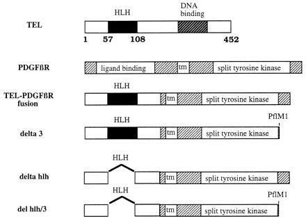
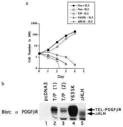
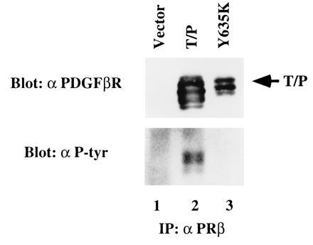
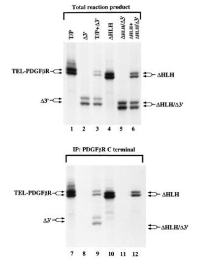
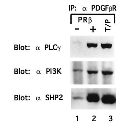
The TEL/PDGF beta R fusion protein is the product of the t(5;12) translocation in patients with chronic myelomonocytic leukemia. The TEL/PDGF beta R is an unusual fusion of a putative transcription factor, TEL, to a receptor tyrosine kinase. The translocation fuses the amino terminus of TEL, containing the helix-loop-helix (HLH) domain, to the transmembrane and cytoplasmic domain of the PDGF beta R. We hypothesized that TEL/PDGF beta R self-association, mediated by the HLH domain of TEL, would lead to constitutive activation of the PDGF beta R tyrosine kinase domain and cellular transformation. Analysis of in vitro-translated TEL/ PDGF beta R confirmed that the protein self-associated and that self-association was abrogated by deletion of 51 aa within the TEL HLH domain. In vivo, TEL/PDGF beta R was detected as a 100-kDa protein that was constitutively phosphorylated on tyrosine and transformed the murine hematopoietic cell line Ba/F3 to interleukin 3 growth factor independence. Transformation of Ba/F3 cells required the HLH domain of TEL and the kinase activity of the PDGF beta R portion of the fusion protein. Immunoblotting demonstrated that TEL/PDGF beta R associated with multiple signaling molecules known to associate with the activated PDGF beta R, including phospholipase C gamma 1, SHP2, and phosphoinositol-3-kinase. TEL/PDGF beta R is a novel transforming protein that self-associates and activates PDGF beta R-dependent signaling pathways. Oligomerization of TEL/PDGF beta R that is dependent on the TEL HLH domain provides further evidence that the HLH domain, highly conserved among ETS family members, is a self-association motif.


Figures

Figure 1
Figure 1
Schematic representation of TEL, PDGFβR, T/P, and mutations of T/P.
Figure 2
Figure 2
T/P transforms Ba/F3 cells to IL-3 factor independence. (a) Ba/F3 cells were transfected with pcDNA3, pcDNA3 T/P, the kinase-inactive mutant, Y635K, or the ΔHLH mutant and selected for G418 resistance. G418-resistant cells were selected for growth in the absence of IL-3. Neo-resistant (2 × 10⁴ cells; pcDNA3, Y635K, and ΔHLH) or IL-3-independent cells (T/P) were washed free of IL-3 and plated on day 0 in RPMI 1640/10% FCS. Viable cells were counted on each day. (b) Expression of T/P and mutants in Ba/F3 cells. Transfected Ba/F3 cells were lysed in 1% Triton X-100/150 mM NaCl/50 mM Tris, pH 8.0, plus protease inhibitors. Lysates were separated by SDS/PAGE, transferred to nitrocellulose, and blotted with anti-PDGFβR antisera. Lanes 2 and 3 show two separate transfections with wild-type T/P.
Figure 3
Figure 3
T/P is constitutively tyrosine-phosphorylated in Ba/F3 cells. Ba/F3 cells were transfected with pcDNA3 alone (lane 1), T/P (lane 2), or a kinase-inactive mutant with a tyrosine-to-lysine mutation at the site corresponding to Y635 in the human PDGFβR (Y635K; lane 3). Cells were selected in 1 mg/ml of G418, and T/P cells were subsequently selected for growth in the absence of IL-3. Cells were lysed and immunoprecipitated with an antibody to the human PDGFβR (Upstate Biotechnology). Immunoprecipitates were separated by SDS/PAGE, transferred to nitrocellulose, and Western blotted using the indicated antisera and enhanced chemiluminescence (ECL) detection methods. All constructs direct the synthesis of a doublet due to the use of an alternative start site for translation in the TEL portion of the fusion cDNA.
Figure 4
Figure 4
T/P dimerizes in vitro and dimerization requires the HLH domain. cDNA constructs for the indicated mutations of T/P were cloned into the pcDNA3 expression vector, and in vitro transcription/translation was performed according to the manufacturer’s instruction (Promega TNT kit) using radiolabeled [35S]methionine. Quantity of DNA added was adjusted to give approximately equal amounts of each translated protein. For lanes 7–12, one-half of the reaction mixture was removed and immunoprecipitated with an antisera to the carboxyl terminus of human PDGFβR. Total reaction products or total immunoprecipitates were separated by SDS/PAGE. The gel was fixed, treated with Enhance (Amersham), dried, and exposed to film at −70° for 30 min.
Figure 5
Figure 5
T/P associates with signaling molecules. Ba/F3–PDGFβR and Ba/F3 T/P cells were lysed and immunoprecipitated with anti-PDGFβR antisera. Immunoprecipitates were separated by SDS/PAGE, transferred to nitrocellulose, and blotted with the indicated antibodies. Ba/F3–PDGFβR were either deprived of IL-3 for 4 hr (−) or stimulated with 100 ng of recombinant human PDGF B for 10 min at 37° (+).

References

    1. Claesson-Welsh L. J Biol Chem. 1994;269:32023–32026. - PubMed
    1. Golub T R, Barker G F, Bohlander S K, Hiebert S W, Ward D C, Bray-Ward P, Morgan E, Raimondi S C, Rowley J D, Gilliland D G G. Proc Natl Acad Sci USA. 1995;92:4917–4921. - PMC - PubMed
    1. Shurtleff S, Buijs A, Behm F, Rubnitz J, Raimondi S, Hancock M, Chan G, Pui C, Grosveld G, Downing J. Leukemia. 1995;9:1985–1989. - PubMed
    1. Papadopoulos P, Ridge S A, Boucher C A, Stocking C, Wiedemann L M. Cancer Res. 1995;55:34–38. - PubMed
    1. Buijs A, Sherr S, van Baal S, van Bezouw S, van der Plas D, van Kessel A D, Riegman P, Deprez R L, Zwarthoff E, Hagemeijer A, Grosveld G. Oncogene. 1995;10:1511–1519. - PubMed

Publication types

MeSH terms

Substances