. 1996 Dec 10;93(25):14857-61.
doi: 10.1073/pnas.93.25.14857.

Effects of protein calorie malnutrition on tuberculosis in mice

Affiliations

Effects of protein calorie malnutrition on tuberculosis in mice

J Chan et al. Proc Natl Acad Sci U S A. .

Abstract

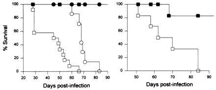
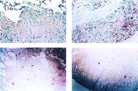
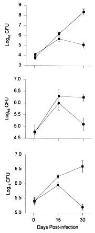
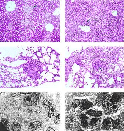
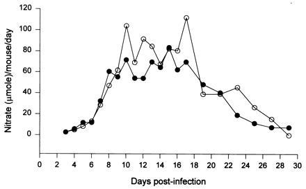
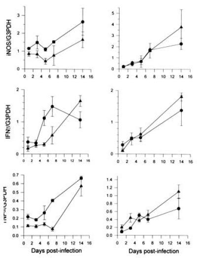
Infectious diseases and malnutrition represent major burdens afflicting millions of people in developing countries. Both conditions affect individuals in industrialized nations, particularly the aged, the HIV-infected, and people with chronic diseases. While malnutrition is known to induce a state of immunodeficiency, the mechanisms responsible for compromised antimicrobial resistance in malnourished hosts remain obscure. In the present study, mice fed a 2% protein diet and developing protein calorie malnutrition, in contrast to well-nourished controls receiving a 20% protein diet, rapidly succumbed to infection with Mycobacterium tuberculosis. Malnourished mice exhibited a tissue-specific diminution in the expression of interferon gamma, tumor necrosis factor alpha, and the inducible form of nitric oxide synthase in the lungs, but not the liver. The expression of these molecules critical to the production of mycobactericidal nitrogen oxides was depressed in malnourished animals in the lungs specifically at early times (< 14 days) after infection. At later times, levels of expression became comparable to those in well-nourished controls, although the bacillary burden in the malnourished animals continued to rise. Nevertheless, urinary and serum nitrate contents, an index of total nitric oxide (NO) production in vivo, were not detectably diminished in malnourished, mycobacteria-infected mice. In contrast to the selective and early reduction of lymphokines and the inducible form of nitric oxide synthase in the lung, a marked diminution of the granulomatous reaction was observed in malnourished mice throughout the entire course of infection in all tissues examined (lungs, liver, and spleen). Remarkably, the progressively fatal course of tuberculosis observed in the malnourished mice could be reversed by restoring a full protein (20%) diet. The results indicate that protein calorie malnutrition selectively compromises several components of the cellular immune response that are important for containing and restricting tuberculous infection, and suggest that malnutrition-induced susceptibility to some infectious diseases can be reversed or ameliorated by nutritional intervention.


Figures

Figure 1
Figure 1
(Left) CD1 mice with PCM (2% protein diet) infected with 106 (□; n = 12 mice) or 104 (○; n = 7 mice) virulent Erdman strain of M. tuberculosis experienced exacerbated course of tuberculous infection compared with controls fed a full protein diet [▪ (n = 9 mice) and • (n = 7 mice) represent control animals infected with 106 and 104 bacilli, respectively]. (Right) Reinstating a full protein diet to M. tuberculosis-infected CD1 mice with PCM reverses the fulminant course of tuberculosis associated with malnutrition. Fourteen days after initiation of the diet, mice fed a protein-deficient diet (2% protein; ▪) or a regular diet (20% protein; □) were infected with 106 virulent Erdman strain of M. tuberculosis. At 18 days postinfection, the diets were switched between the two groups. Seven mice per group were studied.
Figure 2
Figure 2
Immunohistopathological studies of tissues of M. tuberculosis-infected CD1 mice fed a 20% (Left) or 2% (Right) protein diet. Kinyoun’s acid fast stain of lung tissues (Upper) reveals markedly increased bacillary loads in mice with PCM (25 days postinfection) compared with those of controls. Difference in tissue bacillary loads is observed as early as 14 days postinfection in the lungs. Studies of hepatic tissues demonstrated similar findings. Immunocytochemical staining of lung tissues (2.5×) using anti-iNOS antibodies (Lower) demonstrates deficient expression of macrophage iNOS in the lungs of M. tuberculosis-infected mice with PCM. Sections represent tissues obtained from mice 10 days postinfection.
Figure 3
Figure 3
PCM resulted in increased bacillary burden in tissues of CD1 mice infected with 106 virulent Erdman strain of M. tuberculosis. Animals were infected 14 days after initiation of a 2% (▪) or 20% (•) protein diet. Three animals were studied per group per time point. The numbers of cfu shown represent that from the left upper lobe of the lungs (Top), the caudate quarter of the spleen (Middle), and the right lobe of the liver (Bottom). At 12 h after infection, three mice from each group were killed for quantitating the baseline tissue bacillary loads. Bars represent SD.
Figure 4
Figure 4
Granuloma formation in CD1 mice with PCM (Right) is deficient during tuberculous infection (106 cfu M. tuberculosis i.v.) compared with controls given a full protein diet (Left). (Top) Hematoxylin/eosin (H&E)-stained hepatic tissues from mice 7 days postinfection (×20) showing differences in granulomas. In contrast to the well-organized granulomas (arrow) in mice fed a full protein diet (Top Left), those in animals with PCM (Top Right) contain diffuse cellular infiltrates (arrow). (Middle) Necrosis in granulomas in lung tissues of mice with PCM (H&E stained, 14 days postinfection; ×10): Areas of necrosis (arrow) in granulomas of M. tuberculosis-infected mice with PCM (Middle Right). (Bottom) Organization of granulomas: Electron micrographs of liver tissues from mice fed a regular diet (Bottom Left) at 25 days postinfection reveal granulomas with tightly apposed cellular components, compared with those from mice with PCM (Bottom Right), which are loosely organized with apparent intercellular spaces (arrows).
Figure 5
Figure 5
Urinary nitrate contents of BCG-infected CD1 mice with PCM (○) were not diminished compared with those of controls (•) receiving a full-protein diet. Data shown are derived from 24-h urinary nitrate contents of samples pooled from three mice.
Figure 6
Figure 6
Assessment of the expression of iNOS (Top), IFN-γ (Middle), and TNF-α (Bottom) in the lungs (Left) and liver (Right) of M. tuberculosis-infected CD1 mice given a 2% (▴) or a 20% (•) protein diet demonstrates selective attenuation of expression of these three elements in the pulmonic tissues of malnourished animals. Three mice per group per time point were studied. Error bars represent standard error.

Similar articles

Cited by

References

    1. Helweg-Larsen, P., Hoffmeyer, H., Kerler, J., Thaysen, E. H., Thygesen, P. & Wulff, M. H. (1952) Acta Med. Scand. 144, Suppl. 274, 330–362.
    1. McMurray D N. In: Tuberculosis: Pathogenesis, Protection, and Control. Bloom B R, editor. Washington, DC: Am. Soc. Microbiol.; 1994. pp. 135–147.
    1. Scrimshaw N S, Taylor C E, Gordon J E. Am J Med Sci. 1959;237:367–403. - PubMed
    1. Chandra R K. Am J Clin Nutr. 1991;53:1087–1101. - PubMed
    1. McMurray D N. Prog Food Nutr Sci. 1984;8:193–228. - PubMed

Publication types

Substances

LinkOut - more resources