. 1996 Dec 10;93(25):14901-5.
doi: 10.1073/pnas.93.25.14901.

Expression of galanin and a galanin receptor in several sensory systems and bone anlage of rat embryos

Affiliations

Expression of galanin and a galanin receptor in several sensory systems and bone anlage of rat embryos

Z Q Xu et al. Proc Natl Acad Sci U S A. .

Abstract

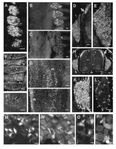
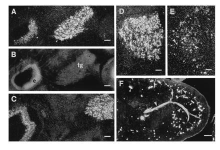
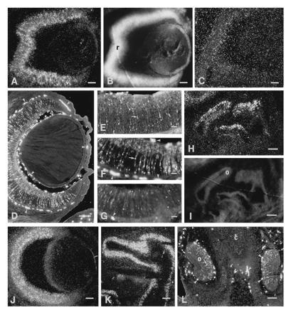
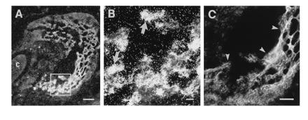
Using in situ hybridization and immunohistochemistry the expression of, respectively, prepro-galanin (pre-pro-GAL) mRNA and GAL receptor-1 mRNA, as well as GAL-like and GAL message-associated peptide-like immunoreactivities, were studied in rats from embryonic day 14 (E14) to postnatal day 1. GAL expression was observed already at E14 in trigeminal and dorsal root ganglion neurons and at E15 in the sensory epithelia in developing ear, eye, and nose, as well as at E19 during bone formation. Also, GAL receptor-1 mRNA was expressed in the sensory ganglia of embryos but appeared later than the ligand. These findings suggest that GAL and/or GAL message-associated peptide may have a developmental role in several sensory systems and during bone formation.


Figures

Figure 1
Figure 1
Darkfield micrographs of DRGs (A–C and F) and spinal cord (G and J) after hybridization with probe complementary to ppGAL mRNA and fluorescence micrographs after incubation with GMAP antiserum (D, E, H, I, K–M, and O). C shows the same section as B stained with bisbenzimide. L shows semiadjacent sections to K incubated with GMAP antiserum preabsorbed with an excess of GMAP. N and P show the same sections as M and O, respectively, after staining with propidium iodide. Distinct ppGAL-mRNA and GMAP expression is seen in DRGs (g) at E15 (A, D, and E), E17 (B, H, and K), and E19 (F). Immunoreactive fibers (arrows) run from the DRGs to the dorsal horn (D, H, and K). GMAP-positive cell bodies are shown in the ventral horn (I). Confocal micrographs show immunoreactivity in mainly the Golgi compartment (arrows) but also as dot-like structures in the thin perinuclear cytoplasm (small arrows) (M–P). The GAL-R1 receptor mRNA is at E17 and is found mainly in the ventral horn (vh) (G) and at E19 in addition in the dorsal horn (dh) (J). Arrowheads in D, H, K, and L point to nonspecific staining, as revealed in the absorption experiments (L). v, vertebra. (AD, FH, and J, bar = 100 μm; I, K, and L, bar = 50 μm; E, bar = 25 μm; M and N, bar = 5 μm; O and P, bar = 2.5 μm.)
Figure 2
Figure 2
Darkfield (A and C–E) and fluorescence micrographs (F) of inner ear (A and C), trigeminal ganglion (A and C–E), and lip (F) showing ppGAL mRNA (A and C–E) and GMAP-LI (F). B shows propidium iodide counterstaining of sections in A. D and E are semiadjacent sections. ppGAL bmRNA expression is seen in the trigeminal ganglion (tg) at E15 (A) and E17 (C and D). GAL-R1 mRNA is present in apparently fewer cells (E). Also, the sensory epithelium (e) of the inner ear shows GAL mRNA expression (A and C). GMAP-LI can be seen in a nerve extending branches in the skin, including into the epithelium (big arrowheads) (F). Small arrowheads point to nonspecific staining. (AC, bar = 100 μm; D and E, 50 μm; F, bar = 200 μm.)
Figure 3
Figure 3
Darkfield (A, C, H, J, and K) and fluorescence (D–G and L) micrographs of the eye (A–G and J) and nasal mucosa (H and K–L) showing ppGAL mRNA (A, H, J, and K) GAL-R1 mRNA (C) and GMAP-LI (D–G and L) expression. B and I show bisbenzimide counterstaining of the same sections as in A and H, respectively. GAL expression (arrows) is seen in the retina (r), with the highest level at E17 (A, D, and F) and lower levels at E15 (E) and E19 (G and J). No distinct GAL-R1 mRNA signal is seen at E17 (C). A strong ppGAL mRNA signal is seen in the dorsal aspects of the nasal mucosa at E15 (H) and E21 (K). GMAP-LI (arrow) is seen in olfactory epithelium (o) (L). c, cartilage. Small arrowheads indicate nonspecific staining. (A–G, J, and K, bar = 100 μm; H, I, and L, bar = 200 μm.)
Figure 4
Figure 4
In situ hybridization autoradiographs of bone formation (A and B). B is a high magnification of A, as indicated by small box. C shows bisbenzimide counterstaining of part of the section shown in A. A strong ppGAL mRNA signal (arrows) is seen in cellular elements (arrowheads) in the zone of bone formation. c, Cartilage. (A, bar = 200 μm; B and C, bar = 50 μm.)

References

    1. Tatemoto K, Rökaeus Å, Jörnvall H, McDonald T J, Mutt V. FEBS Lett. 1983;164:124–128. - PubMed
    1. Rökaeus Å, Melander T, Hökfelt T, Lundberg J M, Tatemoto K, Carlquist M, Mutt V. Neurosci Lett. 1984;47:161–166. - PubMed
    1. Melander T, Hökfelt T, Rökaeus Å. J Comp Neurol. 1986;248:475–517. - PubMed
    1. Skofitsch G, Jacobowitz D M. Peptides. 1985;6:509–546. - PubMed
    1. Ch’ng J L C, Christofides N D, Anand P, Gibson S J, Allen Y S, Su H C, Tatemoto K, Morrison J F B, Polak J M, Bloom S R. Neuroscience. 1985;16:343–354. - PubMed

Publication types

LinkOut - more resources