. 1996 Dec 10;93(25):14950-4.
doi: 10.1073/pnas.93.25.14950.

Down-regulation of transcripts for Na channel alpha-SNS in spinal sensory neurons following axotomy

Affiliations

Down-regulation of transcripts for Na channel alpha-SNS in spinal sensory neurons following axotomy

S Dib-Hajj et al. Proc Natl Acad Sci U S A. .

Abstract

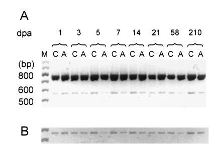
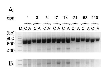
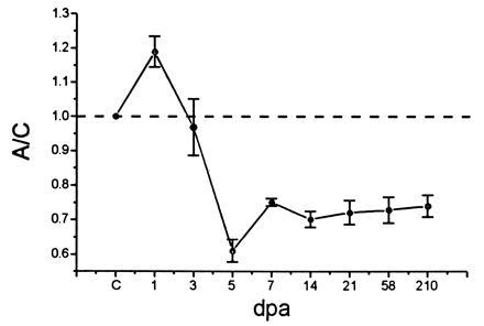
Spinal sensory (dorsal root ganglion; DRG) neurons display slowly inactivating, tetrodotoxin-resistant (TTX-R), and rapidly inactivating, TTX-sensitive (TTX-S) Na currents. Attenuation of the TTX-R Na current and enhancement of TTX-S Na current have been demonstrated in cutaneous afferent DRG neurons in the adult rat after axotomy and may underlie abnormal bursting. We show here that steady-state levels of transcripts encoding the alpha-SNS subunit, which is associated with a slowly inactivating, TTX-R current when expressed in oocytes, are reduced significantly 5 days following axotomy of DRG neurons, and continue to be expressed at reduced levels, even after 210 days. Steady-state levels of alpha-III transcripts, which are present at low levels in control DRG neurons, show a pattern of transiently increased expression. In situ hybridization using alpha-SNS- and alpha-III-specific riboprobes showed a decreased signal for alpha-SNS, and an increased signal for alpha-III, in both large and small DRG neurons following axotomy. Reduced levels of alpha-SNS may explain the selective loss of slowly inactivating, TTX-R current. The abnormal electrophysiological properties of DRG neurons following axonal injury thus appear to reflect a switch in Na channel gene expression.


Figures

Figure 1
Figure 1
RT-PCR coamplification of α-SNS and β-actin transcripts from DRG following unilateral sciatic axotomy. (A) RT-PCR products from control (C) and axotomized (A) DRG at various dpa. Two products can be observed migrating slightly slower than the 800-bp marker (predicted β-actin product: 764 bp) and the 600-bp marker (predicted α-SNS product: 572 bp), respectively. Lane M contains a 100-bp standard. The gel image was digitized, inverted, and printed in black and white sublimation mode. (B) Computer enhancement for photographic purposes of the section of A containing the α-SNS amplification product.
Figure 3
Figure 3
RT-PCR coamplification of α-III and β-actin transcripts. (A) RT-PCR products of transcripts from control (C) and axotomized (A) DRG at various dpa. Two products can be observed, one migrating slower than the 800-bp marker (predicted β-actin product: 764 bp) and another comigrating with the 400-bp marker (predicted α-III product: 412 bp). Lane M, 100-bp standard. (B) Computer enhancement of α-III amplification product.
Figure 2
Figure 2
α-SNS transcript levels following axotomy. Levels of α-SNS transcripts in axotomized (A) and control (C) DRG were normalized to the coamplified β-actin transcripts. The A/C ratio is shown at various dpa. The A/C ratio of α-SNS transcripts in uninjured animals (indicated by C on the abscissa) is set at 1 and indicated by the dashed line. Four animals were used in the analysis at 5, 7, and 21 dpa, while two animals were used for each of the remaining time points. Mean ± SE from at least three independent amplifications was used to calculate each point, employing the origin program.
Figure 4
Figure 4
Sodium channel mRNA α-SNS and α-III expression in control and axotomized DRG neurons. (a) α-SNS is expressed in most small DRG neurons and in some large neurons in control DRG. (b) At 5 dpa, the α-SNS hybridization signal is attenuated. (c) α-III, control DRG. (d) α-III, 5 dpa. (e) Representative field from control DRG hybridized with α-SNS probe, to show + (1), ++ (2), and +++ (3) signals. (×250; bar = 40 μm.)

References

    1. Kostyuk P G, Veselovsky N, Tsyandryenko A. Neuroscience. 1981;6:2423–2430. - PubMed
    1. Yoshida S, Matsuda Y, Samejima A. J Neurophysiol. 1978;639:125–134. - PubMed
    1. Roy M L, Narahashi T. J Neurosci. 1992;12:2104–2111. - PMC - PubMed
    1. Caffrey J M, Eng D L, Black J A, Waxman S G, Kocsis J D. Brain Res. 1992;592:283–297. - PubMed
    1. Elliott A A, Elliott J R. J Physiol (London) 1993;463:39–56. - PMC - PubMed

Publication types

Substances

Associated data

LinkOut - more resources