. 1996 Dec 1;184(6):2091-9.
doi: 10.1084/jem.184.6.2091.

Lineage commitment in the thymus: only the most differentiated (TCRhibcl-2hi) subset of CD4+CD8+ thymocytes has selectively terminated CD4 or CD8 synthesis

Affiliations

Lineage commitment in the thymus: only the most differentiated (TCRhibcl-2hi) subset of CD4+CD8+ thymocytes has selectively terminated CD4 or CD8 synthesis

J A Punt et al. J Exp Med. .

Abstract

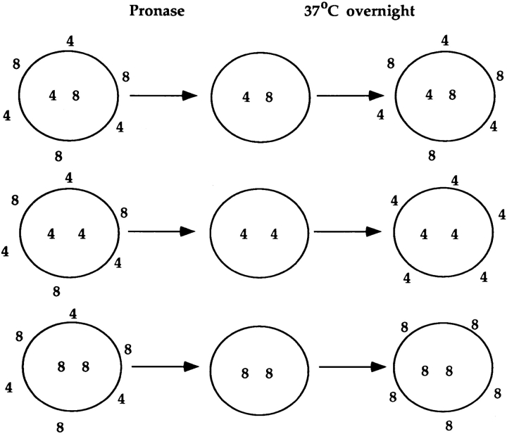
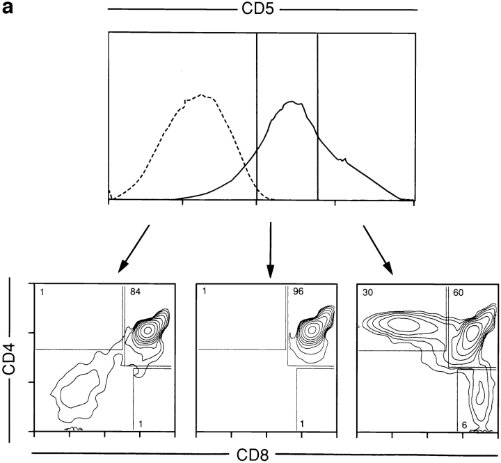
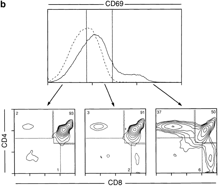
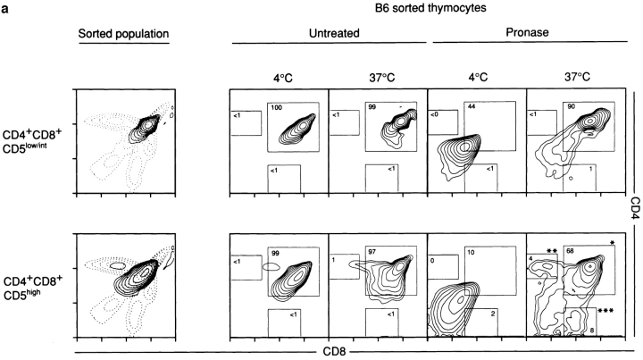
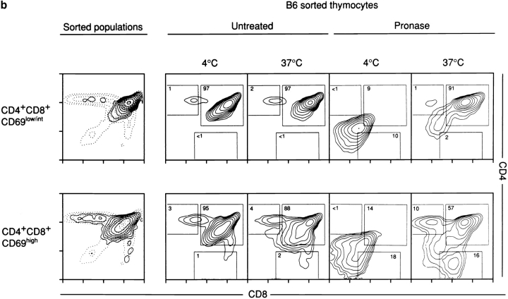
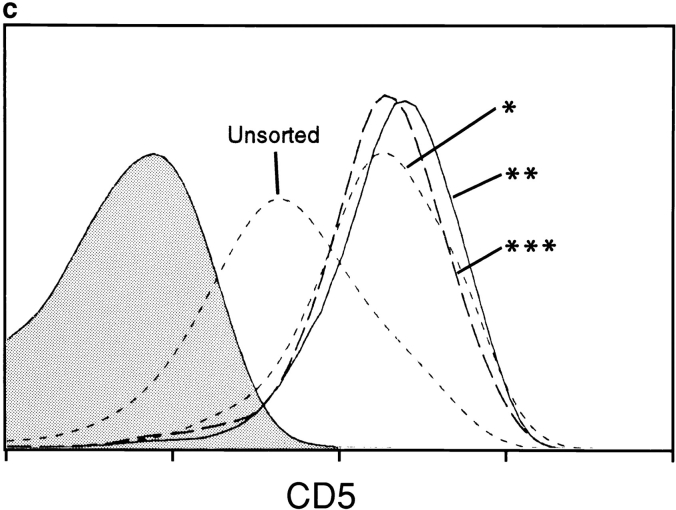
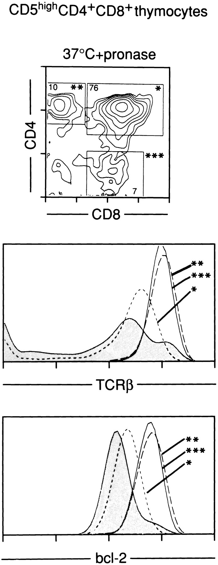
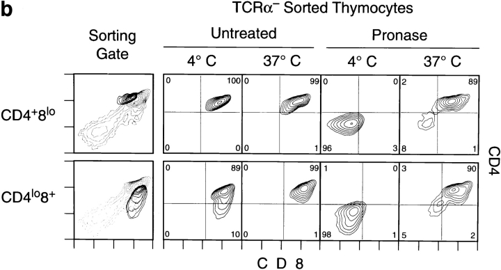
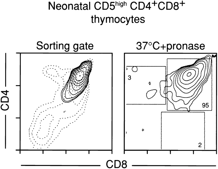
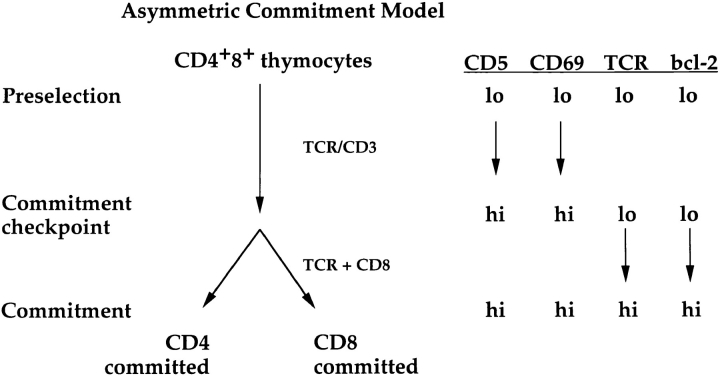
Lineage commitment is a developmental process by which individual CD4+CD8+ (double positive, DP) thymocytes make a decision to differentiate into either CD4+ or CD8+ T cells. However, the molecular event(s) that defines lineage commitment is controversial. We have previously proposed that lineage commitment in DP thymocytes can be molecularly defined as the selective termination of CD4 or CD8 coreceptor synthesis. The present study supports such a molecular definition by showing that termination of either CD4 or CD8 synthesis is a highly regulated event that is only evident within the most differentiated DP subset (CD5hiCD69hiTCRhibcl-2hi). In fact, essentially all cells within this DP subset actively synthesize only one coreceptor molecule. In addition, the present results identify three distinct sub-populations of DP thymocytes that define the developmental progression of the lineage commitment process and demonstrate that lineage commitment is coincident with upregulation of TCR and bcl-2. Thus, this study supports a molecular definition of lineage commitment and uniquely identifies TCRhibcl-2hi DP thymocytes as cells that are already committed to either the CD4 or CD8 T cell lineage.


Figures

Figure 1
Figure 1
Coreceptor reexpression assay. Thymocytes that express both CD4 and CD8 on their surfaces may no longer be actively synthesizing both CD4 and CD8 coreceptor molecules. To reveal the coreceptor(s) that individual CD4+CD8+ thymocytes are actively synthesizing, thymocytes are treated with low concentrations of the protease pronase which strips the surface of the cell of existing CD4 and CD8 protein molecules. Cells are then allowed to reexpress the coreceptor proteins they are actively synthesizing during overnight culture at 37°C. In the schematic, surface CD4 and CD8 proteins are indicated by 4 or 8 outside the circle, and internal CD4 and CD8 mRNAs are indicated by 4 or 8 inside the circle.
Figure 2
Figure 2
CD4+CD8+ thymocytes are heterogeneous for expression of CD5 and CD69. Freshly isolated thymocytes from B6 mice were analyzed by three color flow cytometry for surface expression of CD4, CD8 and CD5 (a) or CD69 (b). Gates subdividing cells into low, intermediate and high expression of CD5 (a) or CD69 (b) are indicated. Contour plots displaying CD4 and CD8 expression of cells in each gate are shown below each gate and the frequency of cells falling into CD4+CD8+, CD4CD8+ and CD4+CD8 subpopulations are indicated. Solid line indicates CD5 or CD69 staining and dashed line indicates negative control (Leu 4) staining.
Figure 2
Figure 2
CD4+CD8+ thymocytes are heterogeneous for expression of CD5 and CD69. Freshly isolated thymocytes from B6 mice were analyzed by three color flow cytometry for surface expression of CD4, CD8 and CD5 (a) or CD69 (b). Gates subdividing cells into low, intermediate and high expression of CD5 (a) or CD69 (b) are indicated. Contour plots displaying CD4 and CD8 expression of cells in each gate are shown below each gate and the frequency of cells falling into CD4+CD8+, CD4CD8+ and CD4+CD8 subpopulations are indicated. Solid line indicates CD5 or CD69 staining and dashed line indicates negative control (Leu 4) staining.
Figure 3
Figure 3
Coreceptor synthesis in CD4+CD8+ thymocytes. Populations of thymocytes from B6 mice were electronically sorted by three color flow cytometry into purified populations of CD5hi versus CD5lo/int CD4+CD8+ cells (a) or CD69hi versus CD69lo/int CD4+CD8+ cells (b). The sorting gates are indicated and the phenotype of the sorted cells after overnight culture at either 4°C or 37°C is shown (left panels). Sorted cells were assessed for coreceptor synthesis using the coreceptor reexpression assay, and the phenotype of pronase stripped cells after overnight culture at either 4°C or 37°C is shown (right panels). The frequency of cells falling into each boxed region is indicated. It is evident after pronase treatment and 37°C culture that CD4+CD8+ thymocytes that express only one coreceptor molecule are present only among the CD5hi (a) and CD69hi (b) CD4+CD8+ thymocyte subpopulations. The CD5 expression levels of sorted CD5hi CD4+CD8+ cells from (a) which only expressed CD8 (***), CD4 (**) or which continued to synthesize both coreceptors (*) were compared (c). Staining with a negative control antibody on unsorted populations is shown as a shaded histogram. This staining shows that the populations continuing to synthesize both CD4 and CD8 could not be distinguished from cells that had selectively terminated CD4 or CD8 synthesis by CD5 expression levels.
Figure 3
Figure 3
Coreceptor synthesis in CD4+CD8+ thymocytes. Populations of thymocytes from B6 mice were electronically sorted by three color flow cytometry into purified populations of CD5hi versus CD5lo/int CD4+CD8+ cells (a) or CD69hi versus CD69lo/int CD4+CD8+ cells (b). The sorting gates are indicated and the phenotype of the sorted cells after overnight culture at either 4°C or 37°C is shown (left panels). Sorted cells were assessed for coreceptor synthesis using the coreceptor reexpression assay, and the phenotype of pronase stripped cells after overnight culture at either 4°C or 37°C is shown (right panels). The frequency of cells falling into each boxed region is indicated. It is evident after pronase treatment and 37°C culture that CD4+CD8+ thymocytes that express only one coreceptor molecule are present only among the CD5hi (a) and CD69hi (b) CD4+CD8+ thymocyte subpopulations. The CD5 expression levels of sorted CD5hi CD4+CD8+ cells from (a) which only expressed CD8 (***), CD4 (**) or which continued to synthesize both coreceptors (*) were compared (c). Staining with a negative control antibody on unsorted populations is shown as a shaded histogram. This staining shows that the populations continuing to synthesize both CD4 and CD8 could not be distinguished from cells that had selectively terminated CD4 or CD8 synthesis by CD5 expression levels.
Figure 4
Figure 4
TCRβ and bcl-2 expression identify CD4+CD8+ thymocytes that have selectively terminated synthesis of one coreceptor molecule. Purified CD5hi CD4+CD8+ thymocytes were obtained by electronic sorting and assessed by the coreceptor reexpression assay. The CD4 and CD8 profile of sorted CD5hi CD4+CD8+ cells treated with pronase and cultured overnight at 37°C is shown as a contour plot. TCRβ and bcl-2 expression of sorted cells which only reexpressed CD8 (***), CD4 (**) or which continued to synthesize both CD4 and CD8 (*) are displayed. The shaded profiles represent staining of unsorted whole thymocytes that had been similarly pronase treated and cultured at 37°C overnight.
Figure 5
Figure 5
Selective termination of coreceptor synthesis requires TCRαβ/CD3 expression. (A) CD5 expression of CD4+CD8+ thymocytes from wild-type and TCRα deficient mice was assessed by flow cytometry. It is evident that the CD5hi CD4+CD8+ thymocyte subpopulation is absent in TCRα-deficient mice. (B) CD4+CD8lo and CD4lo CD8+ subpopulations of thymocytes from a TCRα-deficient mouse were electronically sorted according to the gates indicated and were assessed for coreceptor synthesis by the coreceptor reexpression assay. Frequency of cells falling into each quadrant are indicated. No cells that had selectively terminated CD4 or CD8 synthesis are evident in either population.
Figure 5
Figure 5
Selective termination of coreceptor synthesis requires TCRαβ/CD3 expression. (A) CD5 expression of CD4+CD8+ thymocytes from wild-type and TCRα deficient mice was assessed by flow cytometry. It is evident that the CD5hi CD4+CD8+ thymocyte subpopulation is absent in TCRα-deficient mice. (B) CD4+CD8lo and CD4lo CD8+ subpopulations of thymocytes from a TCRα-deficient mouse were electronically sorted according to the gates indicated and were assessed for coreceptor synthesis by the coreceptor reexpression assay. Frequency of cells falling into each quadrant are indicated. No cells that had selectively terminated CD4 or CD8 synthesis are evident in either population.
Figure 6
Figure 6
CD5hi CD4+CD8+ cells within a newborn thymus have not yet selectively terminated CD4 or CD8 synthesis. Neonatal thymi were harvested within 12 h of birth and CD5hi CD4+CD8+ cells were electronically sorted according to the gate indicated in the left panel. The coreceptor reexpression assay was performed on the sorted subpopulation and the frequency of cells falling into each quadrant is indicated. It is evident that the vast majority of CD5hi CD4+CD8+ thymocytes continued to synthesize both CD4 and CD8; no discrete populations of cells that had selectively terminated CD8 or CD4 synthesis was evident. (For comparison, Fig. 4 displays an experiment performed with CD5hi CD4+CD8+ thymocytes from adult mice.)
Figure 7
Figure 7
Subpopulations of CD4+CD8+ thymocytes, their commitment status, and their relationship to the Asymmetric Commitment Model (12). The results of the present study provide a phenotype that can be assigned to each of the commitment stages proposed in the asymmetric commitment model (12). Preselection CD4+CD8+ thymocytes are too immature to undergo lineage commitment and express low levels of CD5, CD69, TCR and bcl-2. Signals transduced by the TCR/CD3 complex (as result of engagement of those surface molecules that signal through the TCR/CD3 complex, e.g. TCR, Thy-1, etc.) increase CD5 and CD69 expression and promote the differentiation of preselection CD4+CD8+ thymocytes to the point that they are able to undergo lineage commitment (commitment checkpoint). At the commitment checkpoint TCR-signaled CD4+CD8+ thymocytes whose TCR and CD8 molecules are coengaged become CD8-committed, whereas TCR-signaled CD4+CD8+ thymocytes whose TCR and CD8 molecules are not coengaged become CD4-committed. Upregulation of TCR and bcl-2 occurs coincident with lineage commitment so that lineage committed thymocytes express TCR and bcl-2 at high levels.

Similar articles

Cited by

References

    1. Robey E, Fowlkes BJ. Selective events in T cell development. Annu Rev Immunol. 1994;12:675–705. - PubMed
    1. Fowlkes BJ, Schweighoffer E. Positive selection of T cells. Curr Opin Immunol. 1995;5:873–879. - PubMed
    1. Von Boehmer H. CD4/CD8 lineage commitment: Back to instruction? . J Exp Med. 1996;183:713–715. - PMC - PubMed
    1. Davis CB, Killeen N, Crooks ME, Raulet D, Littman DR. Evidence for a stochastic mechanism in the differentiation of mature subsets of T lymphocytes. Cell. 1993;73:237–247. - PubMed
    1. Chan SH, Cosgrove D, Waltzinger C, Benoist C, Mathis D. Another view of the selective model of thymocyte selection. Cell. 1993;73:225–236. - PubMed

Publication types

MeSH terms