. 1996 Dec 1;184(6):2119-28.
doi: 10.1084/jem.184.6.2119.

The Ly-49D receptor activates murine natural killer cells

Affiliations

The Ly-49D receptor activates murine natural killer cells

L H Mason et al. J Exp Med. .

Abstract

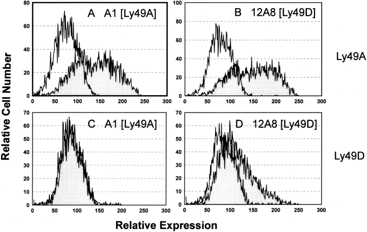
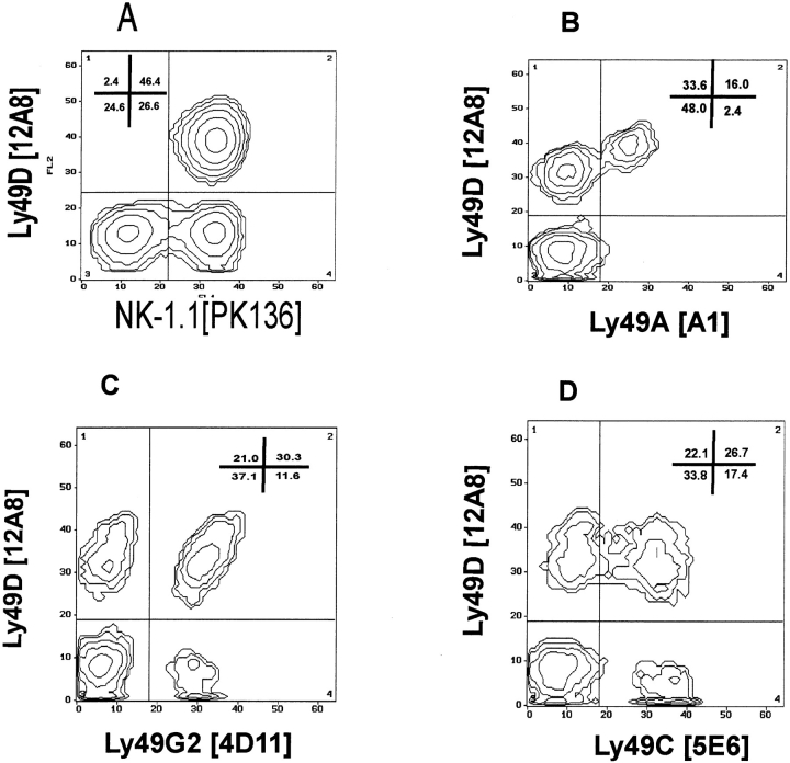
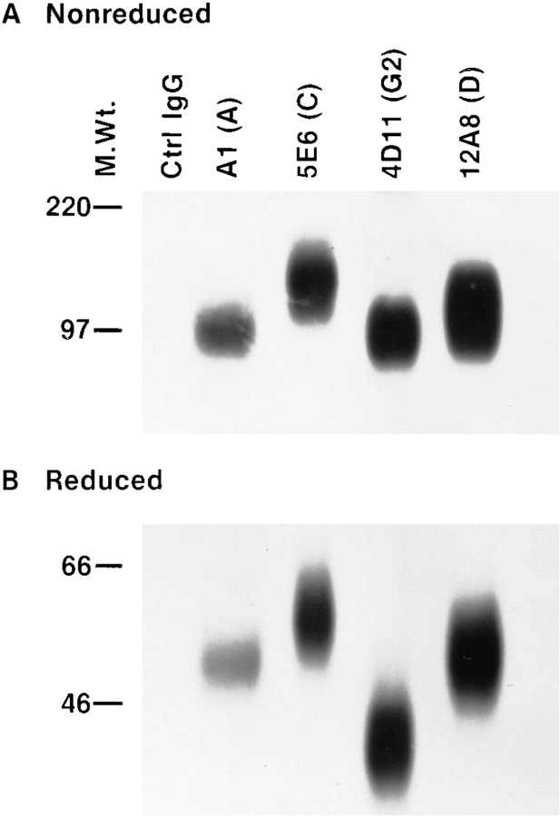
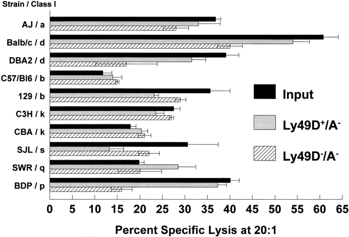
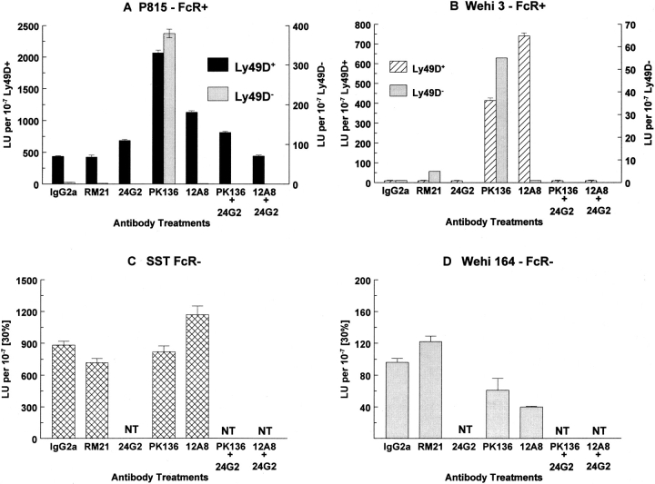
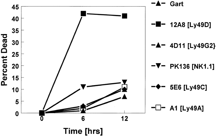
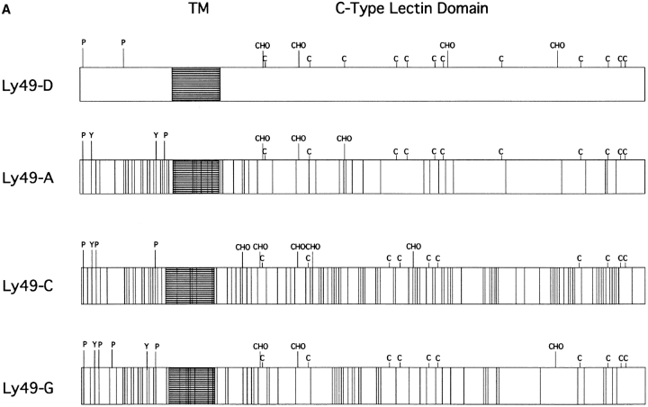
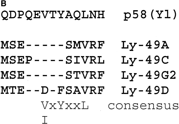
Proteins encoded by members of the Ly-49 gene family are predominantly expressed on murine natural killer (NK) cells. Several members of this gene family have been demonstrated to inhibit NK cell lysis upon recognizing their class I ligands on target cells. In this report, we present data supporting that not all Ly-49 proteins inhibit NK cell function. Our laboratory has generated and characterized a monoclonal antibody (mAb) (12A8) that can be used to recognize the Ly-49D subset of murine NK cells. Transfection of Cos-7 cells with known members of the Ly-49 gene family revealed that 12A8 recognizes Ly-49D, but also cross-reacts with the Ly-49A protein on B6 NK cells. In addition, 12A8 demonstrates reactivity by both immuno-precipitation and two-color flow cytometry analysis with an NK cell subset that is distinct from those expressing Ly-49A, C, or G2. An Ly-49D+ subset of NK cells that did not express Ly-49A, C, and G2 was isolated and examined for their functional capabilities. Tumor targets and concanovalin A (ConA) lymphoblasts from a variety of H2 haplotypes were examined for their susceptibility to lysis by Ly-49D+ NK cells. None of the major histocompatibility complex class I-bearing targets inhibited lysis of Ly-49D+ NK cells. More importantly, we demonstrate that the addition of mAb 12A8 to Ly-49D+ NK cells can augment lysis of Fc gamma R+ target cells in a reverse antibody-dependent cellular cytotoxicity-type assay and induces apoptosis in Ly-49D+ NK cells. Furthermore, the cytoplasmic domain of Ly-49D does not contain the V/Ix-YxxL immunoreceptor tyrosine-based inhibitory motif found in Ly-49A, C, or G2 that has been characterized in the human p58 killer inhibitory receptors. Therefore, Ly-49D is the first member of the Ly-49 family characterized as transmitting positive signals to NK cells, rather than inhibiting NK cell function.


Figures

Figure 2
Figure 2
mAb 12A8 stains Cos-7 cells transfected with Ly-49A and Ly-49D. Cos-7 cells were transfected with either Ly-49A or Ly-49D using Lipofectamine and were harvested after a 72-h culture at 37°C for protein expression. Cells transfected with Ly-49A (A and B) and Ly-49D (C and D) were stained with mAb A1 (A and C) or mAb 12A8 (B and D). mAb 12A8 staining was observed on both Ly-49A and Ly-49D transfectants, whereas mAb A1 stained only Cos-7 cells tranfected with Ly-49A. The unshaded histogram represents staining with FITC-GAM (mAb A1) or FITC-GART (mAb 12A8). Shaded histograms represent staining observed with a primary antibody (A1 or 12A8) plus FITC-GAM or FITC-GART.
Figure 1
Figure 1
mAb 12A8 identifies a distinct subset of C57BL/6 NK cells. Freshly isolated C57BL/6 splenic NK cells were enriched by passage over nylon wool columns, depleted of T and B cells, and prepared for FCA as follows: (A) PK136 plus biotinylated 12A8 followed by FITC– goat anti–mouse IgG2a and Steptavidin-PE, (B) FITC-A1 plus biotinylated 12A8 followed by Streptavidin PE, (C) FITC4D11 plus biotinylated 12A8 followed by Streptavidin PE, and (D) FITC-5E6 plus biotinylated 12A8 followed by Streptavidin PE.
Figure 3
Figure 3
Immunoprecipitation using mAb 12A8 reveals a unique disulfide-linked homodimer. B6 NK cells were cultured for 7 d in high dose IL-2. Cells were radiolabeled with 125I and lysed in 0.5% Triton X-100. Sequential IP was performed on a single lysate by the following mAb crosslinked to protein G–Sepharose: Control rat IgG2A, A1, 5E6, 4D11, and 12A8, respectively. Approximately equal CPMs were applied to a 10% SDSPAGE gel and electrophoresed under both nonreduced (A) and reduced (B) conditions.
Figure 4
Figure 4
Ly-49D+/A and Ly-49D/A NK cells lyse Con A blasts of multiple H-2 haplotypes. Ly49D+/A NK cells were sorted from Ly-49D/A NK cells after partial depletion of the Ly-49C+/A+ and G2+ cells, as described in Materials and Methods. Cells were cultured for 6 d in high dose IL- 2. Con A lymphoblasts were prepared from a variety of H-2 haplotypes representing H-2a, H-2d, H- 2k, H-2s, H-2a, and H-2q. Blasts were labeled with 51Cr and used as targets in a 4-h cytotoxicity assay. E/T ratios of 20:1, 7:1, 2:1, and 0.7:1 were assayed, and the percent of specific lysis at 20:1 is shown. Spontaneous release of targets varied from 19 to 35%. Results also are presented for the input population that was partially depleted of Ly-49A, C, and G2+ cells. The resulting Ly-49 phenotypes of these populations are the same as in the legend to Table 1. These results represent one of two such experiments performed.
Figure 5
Figure 5
mAb 12A8 mediates RADCC of FcγR+ target cells by Ly-49D+, but not Ly-49D, NK cells. Splenic NK cells were depleted of cells expressing Ly-49A, C, and G2. Ly-49D+ and Ly-49D cells were sorted (using biotinylated 12A8 followed by streptavidin PE) into populations that were >98% 12A8+ (Ly-49D+) or 12A8 (Ly-49D). These subsets were cultured in high dose IL-2 for 9 d, washed, and tested against the indicated targets in a 4-h 51Cr-release assay at E/T ratios starting at 10:1. mAbs were added at a final concentration of 2 μg/well. Data are presented as lytic units per 107 cells at 30% lysis. This experiment is one of at least three representative assays (NT, not tested). Both the Ly-49D+ and Ly-49D populations were <5% Ly49A, C, and G2+ at the time of testing.
Figure 6
Figure 6
mAb 12A8 induces apoptosis in Ly-49D+ NK cells. This graph presents the data obtained from a representative experiment in which percent cell death (PI uptake) is calculated after 6 and 12 h of antibody treatment. Cell death is calculated over that observed with media alone (33, 35 & 38% respectively). Maximum cell death was observed after 6 h of treatment with mAb 12A8 and cross-linking. This is a representative experiment of eight similar experiments performed.
Figure 7
Figure 7
(A) Amino acid homology of Ly-49D vs. Ly-49A, C, and G2. This figure is a schematic representation of the proposed amino acid sequences of Ly-49D, A, C, and G2, as described by Smith et al. (3) The highly conserved extracellular cysteine residues are represented (C) along with the proposed glycosylation sites (CHO). Potential serine/threonine phosphorylation sites are labeled in the cytoplasmic region (P). Possible tyrosine phosphorylation sites (Y) are also designated in the cytoplasmic domain. Open spaces in these schematics represent areas of homology between these four proteins, whereas the perpendicular lines shown in Ly-49A, C, and G2 represent individual amino acid differences compared to Ly-49D. (B) Homologous regions from the intracellular domains of the four Ly-49 molecules compared to the human p58 inhibitory receptor. Dashes represent amino acids identical to the p58 sequence. A consensus sequence (V/IxYxxL) for a possible immunoreceptor tyrosine inhibitory motif found in p58 is also present in Ly-49A, C, and G2, but not in Ly-49D.
Figure 7
Figure 7
(A) Amino acid homology of Ly-49D vs. Ly-49A, C, and G2. This figure is a schematic representation of the proposed amino acid sequences of Ly-49D, A, C, and G2, as described by Smith et al. (3) The highly conserved extracellular cysteine residues are represented (C) along with the proposed glycosylation sites (CHO). Potential serine/threonine phosphorylation sites are labeled in the cytoplasmic region (P). Possible tyrosine phosphorylation sites (Y) are also designated in the cytoplasmic domain. Open spaces in these schematics represent areas of homology between these four proteins, whereas the perpendicular lines shown in Ly-49A, C, and G2 represent individual amino acid differences compared to Ly-49D. (B) Homologous regions from the intracellular domains of the four Ly-49 molecules compared to the human p58 inhibitory receptor. Dashes represent amino acids identical to the p58 sequence. A consensus sequence (V/IxYxxL) for a possible immunoreceptor tyrosine inhibitory motif found in p58 is also present in Ly-49A, C, and G2, but not in Ly-49D.

References

    1. Ljunggren HG, Karre K. In search of the “missing self ”: MHC molecules and NK cell recognition. Immunol Today. 1990;11:237–244. - PubMed
    1. Wong S, Freeman D, Kellcher C, Mager D, Takei F. Ly-49 multigene family. New members of a superfamily of type II-membrane proteins with lectin-like domains. J Immunol. 1991;147:1417–1423. - PubMed
    1. Smith HRC, Karlhofer FM, Yokoyama WM. Ly-49 multigene family expressed by IL-2 activated NK cells. J Immunol. 1994;153:1068–1079. - PubMed
    1. Karlhofer FM, Ribaudo RK, Yokoyama WM. MHC class I alloantigen specificity of Ly-49+IL-2 activated natural killer cells. Nature (Lond) 1992;358:21–22. - PubMed
    1. Kane KP. Ly-49 mediates EL4 lymphoma adhesion to isolated class I major histocompatibility complex molecules. J Exp Med. 1994;179:1011–1015. - PMC - PubMed

MeSH terms