. 1996 Dec 1;184(6):2153-65.
doi: 10.1084/jem.184.6.2153.

HLA-DM interactions with intermediates in HLA-DR maturation and a role for HLA-DM in stabilizing empty HLA-DR molecules

Affiliations

HLA-DM interactions with intermediates in HLA-DR maturation and a role for HLA-DM in stabilizing empty HLA-DR molecules

L K Denzin et al. J Exp Med. .

Abstract

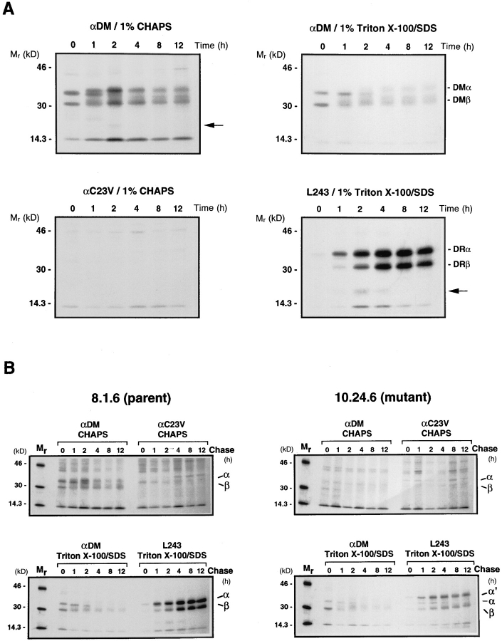
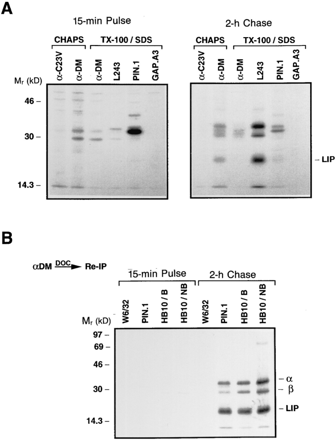
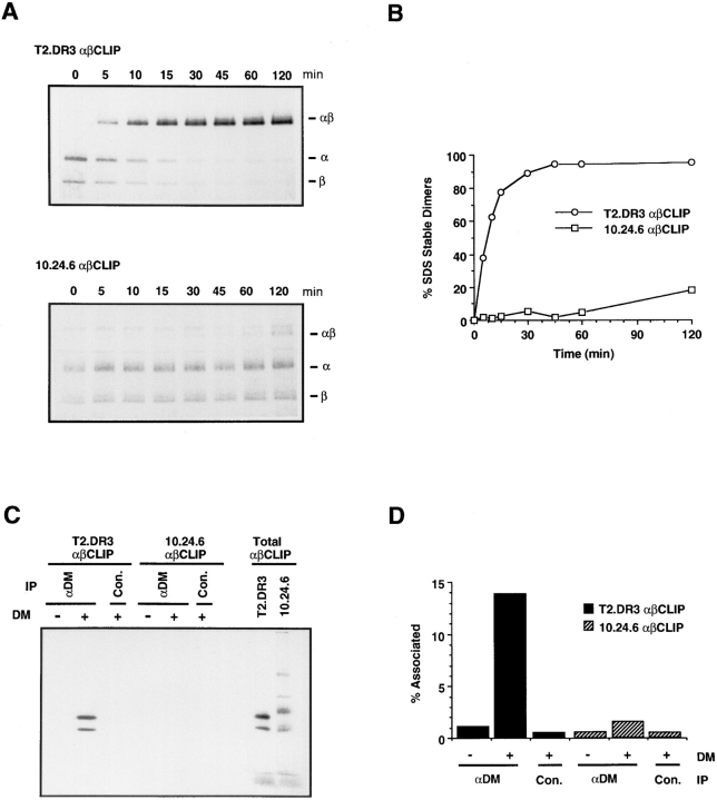
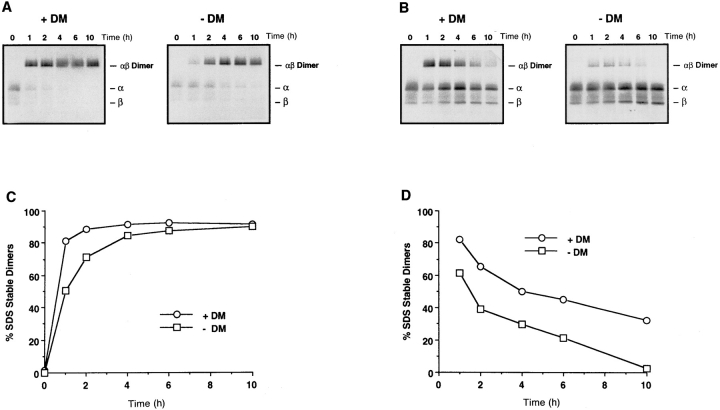
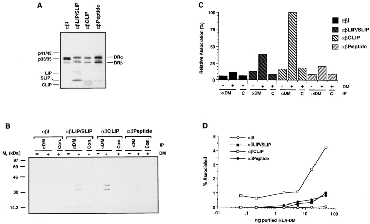
Major histocompatibility complex (MHC) class II-positive cell lines which lack HLA-DM expression accumulate class II molecules associated with residual invariant (I) chain fragments (class II-associated invariant chain peptides [CLIP]). In vitro, HLA-DM catalyzes CLIP dissociation from class II-CLIP complexes, promoting binding of antigenic peptides. Here the physical interaction of HLA-DM with HLA-DR molecules was investigated. HLA-DM complexes with class II molecules were detectable transiently in cells, peaking at the time when the class II molecules entered the MHC class II compartment. HLA-DR alpha beta dimers newly released from I chain, and those associated with I chain fragments, were found to associate with HLA-DM in vivo. Mature, peptide-loaded DR molecules also associated at a low level. These same species, but not DR-I chain complexes, were also shown to bind to purified HLA-DM molecules in vitro. HLA-DM interaction was quantitatively superior with DR molecules isolated in association with CLIP. DM-DR complexes generated by incubating HLA-DM with purified DR alpha beta CLIP contained virtually no associated CLIP, suggesting that this superior interaction reflects a prolonged HLA-DM association with empty class II dimers after CLIP dissociation. Incubation of peptide-free alpha beta dimers in the presence of HLA-DM was found to prolong their ability to bind subsequently added antigenic peptides. Stabilization of empty class II molecules may be an important property of HLA-DM in facilitating antigen processing.


Figures

Figure 1
Figure 1
(A) Kinetics of HLADM and HLA-DR association in wild-type Pala cells. Cells were pulse labeled with [35S]methionine for 15 min and chased in the presence of an excess of methionine and cysteine for the indicated periods of time (h). Class II and DM molecules were immunoprecipitated at each time point from CHAPS or Triton X-100 solubilized lysates (as indicated above each gel) with the DR-specific mAb L243 or with a polyclonal rabbit antiserum specific for the DM heterodimer (αDM) and analyzed by SDSPAGE (10.5%). Control precipitates were performed from CHAPS solubilized lysates with an antiserum specific for an influenza hemagglutinin peptide (αC23V). The bands corresponding to the DMα, DMβ, DRα and DRβ proteins are marked on the right and an I degradation fragment is indicated by a solid arrow head. (B) HLADM does not associate with HLA-DR in 10.24.6 cells expressing a mutant DRα chain containing an extra N-linked glycan. Mutant 10.24.6 and wild-type 8.1.6 cells were pulsed labeled, chased and immunoprecipitated as in A. The positions of the individual class II and DMα and β chains are indicated on the right and α′ marks the position of the mutant DRα chain expressed by 10.24.6 cells.
Figure 2
Figure 2
(A and B) HLA-DM associates with DR in a post-Golgi compartment. Pala cells were pulsed labeled with [35S]methionine for 15 min and chased for the indicated periods of time (h). DM-DR complexes were immunoprecipitated at each time point from CHAPS solubilized lysates with anti-DM (αDM). DM-associated class II molecules were eluted with 0.5% DOC and the supernatants reprecipitated with DR β chain-specific mAb HB10A (A) or control mAb W6/32 (B). The precipitates were either treated with Endo H or mock treated and analyzed by SDS-PAGE (10.5%). Locations of Endo H resistant (R) and sensitive (S) DR α and β bands are indicated on the right. (C) HLA-DM associates with SDS-stable αβ dimers. Pala cells were pulse labeled, immunoprecipitated and re-precipitated with mAb HB10A as in A. After the addition of Laemmli sample buffer, the precipitates were incubated at room temperature for 30 min and analyzed by SDS-PAGE (10.5%). The positions of the individual α and β chains and the αβ dimers are indicated on the right.
Figure 3
Figure 3
HLA-DM associates with αβLIP and αβSLIP complexes. (A) Pala cells were pulse labeled for 15 min with [35S]methionine and chased for 2 h in the presence of an excess of methionine and cysteine. Class II, DM and I chain molecules were immunoprecipitated from CHAPS solubilized lysates with anti-DM (αDM) or control anti-C23V (αC23V) or from Triton X-100 solubilized lysates with anti-DM, DR-specific mAb L243, I chain–specific mAb PIN.1 or control mAb GAP.A3 and analyzed by SDS-PAGE (12%). (B) Wild-type Pala cells were pulse labeled and chased as in A. DM-associated class II and class II–I chain complexes were eluted with 0.5% DOC, the supernatants reprecipitated with mAbs PIN.1, HB10A, and W6/32 (control) and analyzed by SDS-PAGE (12%). The positions of the individual DR α and β chains and LIP are indicated on the right. The right lane shows the HB10A-precipitated material run without boiling the sample.
Figure 4
Figure 4
(A and B) HLADR3 αβCLIP complexes from 10.24.6 cells cannot be loaded by affinity-purified HLA-DM in vitro. Purified HLA-DR3 αβCLIP complexes from T2.DR3 and mutant 10.24.6 cells were incubated with the DR3-restricted MOMP peptide and 2 ng of affinity-purified HLA-DM at 37°C for the indicated times at pH 5.0. After neutralization, samples were analyzed by SDS-PAGE (10.5%) (A) and the percent SDS-stable dimers formed at each time point quantitated by image analysis (B). The positions of the individual α and β chains and αβ dimers are indicated on the right. (C and D) HLA-DM associates with αβCLIP complexes in vitro. Radiolabeled αβCLIP complexes from T2.DR3 and 10.24.6 cells were incubated in the presence (50 ng) or absence of affinitypurified HLA-DM at 37°C for 30 min at pH 5.0. The pH was adjusted to 6.0 and the reaction mixtures were immunoprecipitated with anti-DM (αDM) or control anti-C23V (con.) and analyzed by SDS-PAGE (10.5%) (C) and quantitated by image analysis (D). The lanes labeled Total αβCLIP represent onetenth the amount of radiolabeled αβCLIP used in the reaction mixture and was used to calculate the percentage DM associated class II (% Associated). IP in C, indicates the antibody used for immunoprecipitation.
Figure 5
Figure 5
Stabilization of empty class II molecules by HLA-DM. (A and B) Loading of class II αβCLIP complexes in octyl glucoside versus loading in octyl glucoside and DM. Purified HLA-DR3 αβCLIP complexes (25 nM) from T2.DR3 cells were incubated in 1% octyl glucoside with the DR3-specific MOMP peptide in the presence or absence of 25 nM affinity-purified HLA-DM at 37°C for the indicated times at pH 4.5. After neutralization, samples were analyzed by SDS-PAGE (A) and the percent SDS-stable dimers formed in the presence and absence of HLA-DM at each time point were quantitated by image analysis (B). The positions of the individual α and β chains and the αβ dimers are indicated on the right. (C and D) HLA-DM stabilizes empty class II molecules. Radiolabeled DR3 αβCLIP complexes (25 nM) were incubated at 37°C in 1% octyl glucoside in the presence and absence of HLA-DM (25 nM) for the indicated times at pH 4.5. At each time point, 1 μM MOMP peptide was added and the samples were incubated at 37°C for an additional 45 min. After neutralization, samples were analyzed by SDS-PAGE (C). To quantitate the results in panel C, the amount of SDSstable dimers at each time point is represented in panel D as the percentage of the maximal available SDS-stable dimers generated at the same time point in the presence and absence of DM but with MOMP continuously present (B).
Figure 6
Figure 6
HLA-DM interacts in vitro with αβ dimers associated with I chain fragments or endogenous peptides but not αβI. (A) SDS-PAGE (12%) analysis of radiolabeled, purified αβI, αβLIP/SLIP, αβCLIP, and αβ peptide complexes used for the experiments shown in B and D. The positions of the individual DR α and β chains are indicated on the right and the positions of the I chain (p41/43 and p33/35), LIP, SLIP and CLIP are indicated on the left. (B and C) Radiolabeled complexes (as indicated) were incubated in the presence and absence of purified DM at 37°C for 30 min at pH 5.0, the pH was adjusted to 6.0 and the reaction mixtures were immunoprecipitated with anti-DM (αDM) or control anti-C23V (con.). After analysis of the precipitates by SDS-PAGE (12%) (B), the results were quantitated by image analysis (C) to determine the relative percentage each DR complex that remained associated with DM. (D) Purified complexes were incubated with various concentrations of affinity-purified HLA-DM as indicated (50 ng to 0.156 ng) at 37°C for 30 min at pH 5.0 and the reaction mixtures were immunoprecipitated with anti-DM. After analysis of the precipitates by SDSPAGE, the percentage of each class II complex coprecipitated with DM was determined for each concentration by image analysis.

References

    1. Unanue ER. Antigen-presenting function of the macrophage. Ann Rev Immunol. 1984;2:395–428. - PubMed
    1. Cresswell, P. 1994. Assembly, transport and function of MHC class II molecules. Annu. Rev. Immunol 12:259–293. - PubMed
    1. Jones PP, Murphy DB, Hewgill D, McDevitt HO. Detection of a common polypeptide chain in I-A and I-E sub-region immunoprecipitates. Mol Immunol. 1979;16:51–60. - PubMed
    1. Machamer CE, Cresswell P. Biosynthesis and glycosylation of the invariant chain associated with HLA-DR antigens. J Immunol. 1982;129:2564–2569. - PubMed
    1. Anderson KS, Cresswell P. A role for calnexin (IP90) in the assembly of class II MHC molecules. EMBO (Eur Mol Biol Organ) J. 1994;13:675–682. - PMC - PubMed

Publication types