

Identification of TRP-2 as a human tumor antigen recognized by cytotoxic T lymphocytes
- PMID: 8976176
- PMCID: PMC2211562
- DOI: 10.1084/jem.184.6.2207
Identification of TRP-2 as a human tumor antigen recognized by cytotoxic T lymphocytes
Abstract
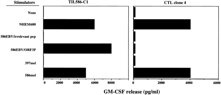
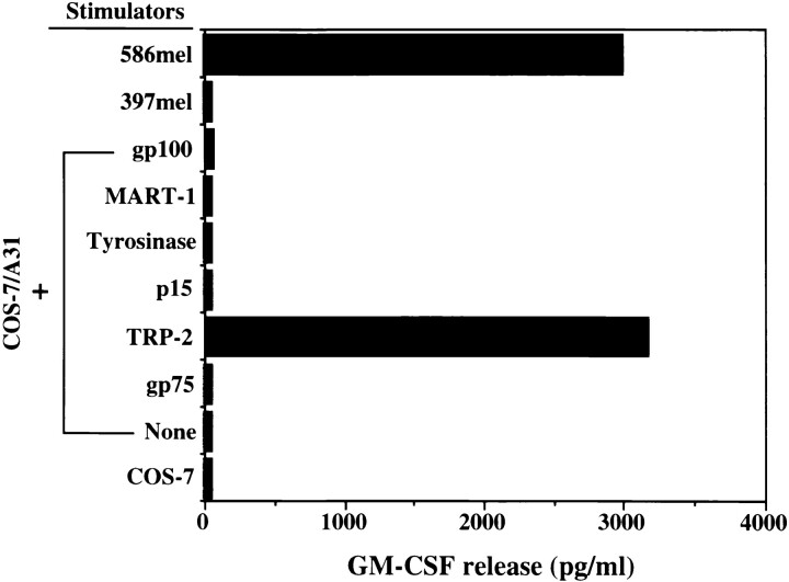
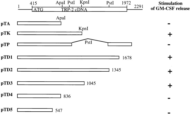
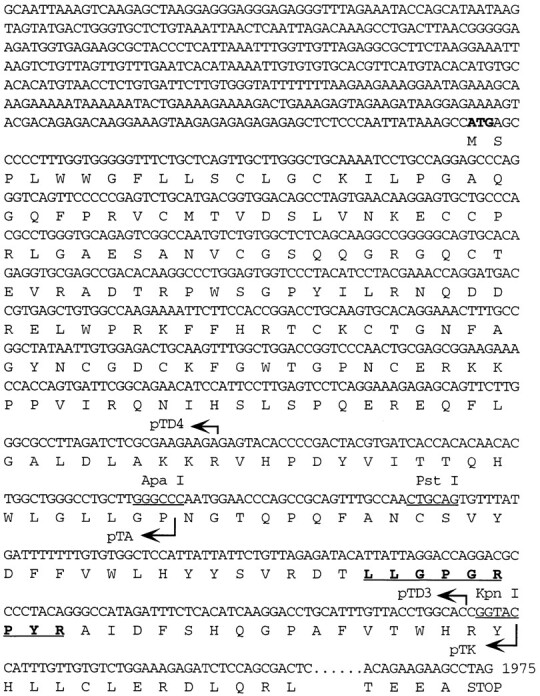
The infusion of TIL586 along with interleukin-2 into the autologous patient with metastatic melanoma resulted in the objective regression of tumor. A gene encoding a tumor antigen recognized by TIL586 was previously isolated and shown to encode gp75 or TRP-1. Here we report that TRP-2 was identified as a second tumor antigen recognized by a HLA-A31-restricted CTL clone derived from the TIL586 cell line. The peptide LLPGGRPYR epitope was subsequently identified from the coding region of TRP-2 based on studies of the recognition of truncated TRP-2 cDNAs and the HLA-A31 binding motif. This epitope peptide was capable of sensitizing target cells for lysis by a CTL clone at 1 nM peptide concentration. Although some modified peptides could be recognized by the CTL clone, none were found to be better recognized by T cells than the parental peptide. Like other melamona differentiation antigens, TRP-2 was only expressed in melanoma, melanocytes, and retina, but not in other human tissues tested.
Figures
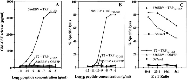
 ) and T2 (non-A31) cells were pulsed with the TRP197–205 (
 ) at various peptide concentrations for 90 min. ORF3P as a control peptide was pulsed onto 586EBV B cells (--▵--). GM-CSF release by CTL clone 4 was determined after coincubation with 586EBV B cells pulsed with TRP197–205 and ORF3P, and T2 cells pulsed with TRP197–205. (B) Sensitization of the target cells for lysis by CTL clone 4 at different peptide concentrations. 586EBV B cells were incubated with TRP197–205 ( ), an irrelevant peptide ORF3P (--▵--), and T2 cells pulsed with TRP197–205 ( ) at various peptide concentrations. After peptide incubation, target cells were labeled for 30 min. Following washes, cytolytic activity of CTL clone 4 at an E/T of 40:1 was measured after a 4 h incubation of T cells with target cells. (C) Lysis of the target cells by CTL clone 4 at different E/T. Target 586EBV cells were separately incubated with TRP197–205 ( ) or the irrelevant peptides ORF3P (--▵--), and target T2 cells were incubated with the TRP197–205 peptide ( ) for 90 min. 586mel (
 ) and 397mel (
 ) were used as positive and negative controls, respectively.
References
-
- Rosenberg SA, Packard BS, Aebersold PM, Solomon D, Topalian SL, Toy ST, Simon P, Lotze MT, Yang JC, Seipp CA, et al. Use of tumor infiltrating lymphocytes and interleukin-2 in the immunotherapy of patients with metastatic melanoma. N Engl J Med. 1988;319:1676–1680. - PubMed
-
- Rosenberg SA, Yannelli JY, Yang JC. Treatment of patients with metastatic melanoma using autologous tumor–infiltrating lymphocytes and interleukin-2. J Natl Cancer Inst. 1994;86:1159–1166. - PubMed
-
- Boon T, Cerottini J-C, Van Den Eynde B, Van der Bruggen P, Van Pel A. Tumor antigens recognized by T lymphocytes. Annu Rev Immunol. 1994;12:337–365. - PubMed
Publication types
MeSH terms
Substances
LinkOut - more resources
Full Text Sources
Other Literature Sources
Medical
Research Materials
