. 1996 Dec 1;184(6):2231-41.
doi: 10.1084/jem.184.6.2231.

Antineutrophil cytoplasmic autoantibodies interact with primary granule constituents on the surface of apoptotic neutrophils in the absence of neutrophil priming

Affiliations

Antineutrophil cytoplasmic autoantibodies interact with primary granule constituents on the surface of apoptotic neutrophils in the absence of neutrophil priming

H M Gilligan et al. J Exp Med. .

Abstract

The pathogenic role of antineutrophil cytoplasmic autoantibodies (ANCA) remains controversial because of the difficulty in explaining how extracellular ANCA can interact with intracellular primary granule constituents. It has been postulated that cytokine priming of neutrophils (PMN), as may occur during a prodromal infection, is an important trigger for mobilization of granules to the cell surface, where they may interact with ANCA. We show by electron microscopy that apoptosis of unprimed PMN is also associated with the translocation of cytoplasmic granules to the cell surface and alignment just beneath an intact cell membrane. Immunofluorescent microscopy and FACS analysis demonstrate reactivity of ANCA-positive sera and antimyeloperoxidase antibodies with apoptotic PMN, but not with viable PMN. Moreover, we show that apoptotic PMN may be divided into two subsets, based on the presence or absence of granular translocation, and that surface immunogold labeling of myeloperoxidase occurs only in the subset of PMN showing translocation. These results provide a novel mechanism that is independent of priming, by which ANCA may gain access to PMN granule components during ANCA-associated vasculitis.


Figures
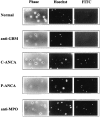
Figure 1
Figure 1
ANCA bind to the surface of unprimed apoptotic PMN. Unprimed apoptotic PMN were treated with polyclonal anti-MPO Ab, normal serum, anti-GBM serum, and high titer C- and P-ANCA sera. Cells were photographed under epifluorescence microscopy for Hoechst, PI, and FITC. All cells excluded PI and so had intact cell membranes. Apoptotic PMN with nuclear condensation comprised two distinct subpopulations with bright or faint Hoechst staining. Surface FITC staining of apoptotic PMN by ANCA sera and anti-MPO Ab occurred more commonly in apoptotic PMN with fainter Hoechst staining.
Figure 2
Figure 2
Anti-MPO Ab stains a subpopulation of unprimed apoptotic PMN. Freshly isolated PMN were treated with anti-MPO Ab (a), P-ANCA serum (b), or C-ANCA serum (c), and were analyzed by FACS® for Hoechst and FITC staining. Also, PMN were cultured overnight to induce apoptosis, treated with antiMPO Ab, and were analyzed by FACS® for Hoechst and FITC staining (d and e). PI-positive cells were excluded from analysis (region defined in 2 d). A minority of PI-positive cells were necrotic (<1% of total PMN), based on increased cell size. The majority had decreased cell size, and were felt to represent postapoptotic cells that had lost membrane integrity. These apoptotic cells were excluded, since loss of membrane integrity might permit binding of ANCA to cytoplasmic primary granules. The remaining cells, including viable and apoptotic PMN, were examined for nuclear staining by Hoechst dye and surface FITC staining by antiMPO Ab or human sera.
Figure 2
Figure 2
Anti-MPO Ab stains a subpopulation of unprimed apoptotic PMN. Freshly isolated PMN were treated with anti-MPO Ab (a), P-ANCA serum (b), or C-ANCA serum (c), and were analyzed by FACS® for Hoechst and FITC staining. Also, PMN were cultured overnight to induce apoptosis, treated with antiMPO Ab, and were analyzed by FACS® for Hoechst and FITC staining (d and e). PI-positive cells were excluded from analysis (region defined in 2 d). A minority of PI-positive cells were necrotic (<1% of total PMN), based on increased cell size. The majority had decreased cell size, and were felt to represent postapoptotic cells that had lost membrane integrity. These apoptotic cells were excluded, since loss of membrane integrity might permit binding of ANCA to cytoplasmic primary granules. The remaining cells, including viable and apoptotic PMN, were examined for nuclear staining by Hoechst dye and surface FITC staining by antiMPO Ab or human sera.
Figure 3
Figure 3
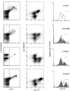
ANCA sera stain a subpopulation of unprimed apoptotic PMN. Apoptotic PMN were treated with anti-MPO Ab, normal serum, antiGBM serum, and C-ANCA and P-ANCA sera. PMN, selected as described in the legend of Fig. 2, were examined for Hoechst and surface FITC staining. Left-hand panels depict scatter plots of Hoechst vs. PI staining. Middle panels are gated to include only PI-negative cells, and depict scatter plots of Hoechst vs. FITC staining. Shaded histograms in the right-hand panels are derived from the scatter plots and depict relative cell number as a function of log FITC fluorescence. Unshaded histograms represent control data from healthy volunteers.
Figure 4
Figure 4
ANCA sera stain a subpopulation of unprimed apoptotic PMN. PMN apoptosis was induced by overnight culture with (a) or without (b) CHX. Scatter plots were gated into the same three subpopulations shown in Fig. 3. Mean channel FITC fluorescence for each subpopulation was compared among normal sera (n = 12), C-ANCA sera (n = 5), and P-ANCA sera (n = 5). PMN were selected as described in the legend of Fig. 2.
Figure 4
Figure 4
ANCA sera stain a subpopulation of unprimed apoptotic PMN. PMN apoptosis was induced by overnight culture with (a) or without (b) CHX. Scatter plots were gated into the same three subpopulations shown in Fig. 3. Mean channel FITC fluorescence for each subpopulation was compared among normal sera (n = 12), C-ANCA sera (n = 5), and P-ANCA sera (n = 5). PMN were selected as described in the legend of Fig. 2.
Figure 5
Figure 5
Surface staining of unprimed apoptotic PMN correlates with the presence of ANCA. PMN apoptosis was induced as described in the legend of Fig. 4. Positively staining PMN were defined as cells whose log-FITC fluorescence was greater than a threshold equal to the mean channel fluorescence with normal human sera for the high-FITC apoptotic population plus five standard deviations (see Fig. 4). Horizontal bars in the right-hand panels of Fig. 3 indicate the regions of positive log FITC fluorescence. PMN staining was compared among normal sera (n = 12), C-ANCA sera (n = 5), and P-ANCA sera (n = 5). PMN were selected as described in the legend of Fig. 2.
Figure 6
Figure 6
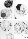
Cytoplasmic granules translocate to the cell surface during apoptosis. PMN, cultured overnight with CHX, were stained with anti-MPO Ab and sorted into two populations by FITC staining. (A) Low FITC staining. Shown are a viable (upper) and an apoptotic PMN (lower). The viable PMN has a multisegmented nucleus (N), many granules throughout the cytoplasm, and an irregular cell outline with membrane ruffling. The apoptotic PMN shows coalescence of the nuclear lobes, preservation of cytoplasmic granules, and a smooth rounded cell outline. (B) High-FITC staining. Shown is an apoptotic PMN sharing many features of apoptosis as within the low-FITC population, except for fewer cytoplasmic granules, many aligned just beneath an intact cell membrane (arrow heads). (C) High FITC staining. Shown are two apoptotic PMN, with nuclear disintegration (*) and decreased numbers of cytoplasmic granules, many again aligned beneath an intact cell membrane. (D) HighFITC staining. Shown is an apoptotic PMN with nuclear disintegration and loss of cell membrane integrity (arrow heads). This PMN is postapoptotic. Original magnifications: (A) ×8,700; (B and C) ×10,800; (D) ×15,000.
Figure 7
Figure 7
PMN apoptosis leads to decreased cellular content of ANCA Ag. PMN were cultured overnight, treated with P-ANCA (a) or C-ANCA (b) sera, and then analyzed by FACS® for PI and FITC staining. All PMN were included.
Figure 7
Figure 7
PMN apoptosis leads to decreased cellular content of ANCA Ag. PMN were cultured overnight, treated with P-ANCA (a) or C-ANCA (b) sera, and then analyzed by FACS® for PI and FITC staining. All PMN were included.
Figure 9
Figure 9
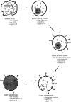
PMN undergoing apoptosis progress through discrete phases. During early apoptosis, nuclear chromatin condenses and nuclear lobes coalesce into a single body. Nucleus disintegration occurs with movement into late apoptosis. Translocation and loss of cytoplasmic primary granules is more distinct during late-phase apoptosis, but also occurs in early apoptotic PMN before nuclear disintegration. ANCA binding correlates with cytoplasmic granular translocation. PMN eventually move into a postapoptotic phase, during which there is loss of cell membrane integrity.
Figure 8
Figure 8
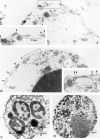
Immunogold labeling localizes MPO on the surface of apoptotic PMN. PMN were cultured overnight with CHX and treated with anti-MPO Ab, followed by gold-conjugated anti–rabbit IgG. To restrict labeling to the PMN surface, fixation occurred after treatment with both Ab. (A and B) Two apoptotic PMN with surface gold labeling. Both show decreased numbers of cytoplasmic granules, many aligned beneath an intact cell membrane (arrow heads). The PMN in B has a coalesced nucleus (N), whereas that in A has undergone nuclear disintegration (*). Boxed areas, showing areas of surface gold labeling, are enlarged within insets. These PMN are representative of the same two phenotypes sorted into the high FITC population for Fig. 6. (C) No surface gold labeling occurs on viable PMN. (D) No surface gold labeling occurs on apoptotic PMN without primary granule translocation. Original magnifications: (A and B) ×35,000; (insets A and B) ×54,000; (C) ×12,300; (D) ×14,800.

Similar articles

Cited by

References

    1. Van der Woude, F.J., N. Rasmussen, S. Lobatto, A. Wiik, H. Permin, L.A. van Es, M. van der Giessen, G.K. van der Hem, and T.H. The. 1985. Autoantibodies against neutrophils and monocytes. Tools for diagnosis and marker of disease activity in Wegener's granulomatosis. Lancet. i:425–429. - PubMed
    1. Falk RJ, Jeannette JC. Anti-neutrophil cytoplasmic autoantibodies with specificity for myeloperoxidase in patients with systemic vasculitis and idiopathic necrotizing and crescentic glomerulonephritis. N Eng J Med. 1988;318:1651–1657. - PubMed
    1. Nölle B, Specks U, Lüdemann J, Rohrbach MS, DeRemee RA, Gross WL. Anticytoplasmic autoantibodies: their immunodiagnostic value in Wegener granulomatosis. Ann Int Med. 1989;111:28–40. - PubMed
    1. Cohen Tervaert, J.W., R. Goldschmeding, J.D. Elema, P.C. Limburg, M. van der Giessen, M.G. Huitema, M.I. Koolen, R.J. Hené, T.H. The, G.K. van der Hem, et al. Association of autoantibodies to myeloperoxidase with different forms of vasculitis. Arthritis Rheum. 1990;33:1264–1272. - PubMed
    1. Lesavre P. Antineutrophil cytoplasmic autoantibodies antigen specificity. Am J Kidney Dis. 1991;18:159–163. - PubMed

Publication types

MeSH terms