. 1996 Dec 1;184(6):2287-300.
doi: 10.1084/jem.184.6.2287.

Cross-linking of Fas by antibodies to a peculiar domain of gp120 V3 loop can enhance T cell apoptosis in HIV-1-infected patients

Affiliations

Cross-linking of Fas by antibodies to a peculiar domain of gp120 V3 loop can enhance T cell apoptosis in HIV-1-infected patients

F Silvestris et al. J Exp Med. .

Abstract

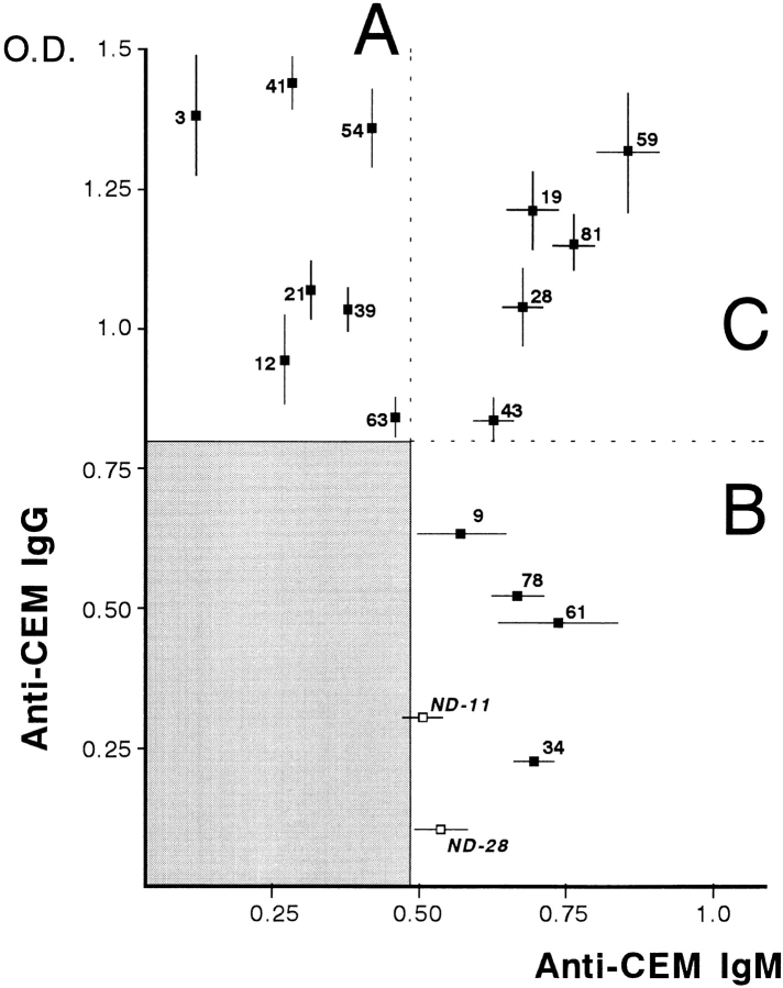
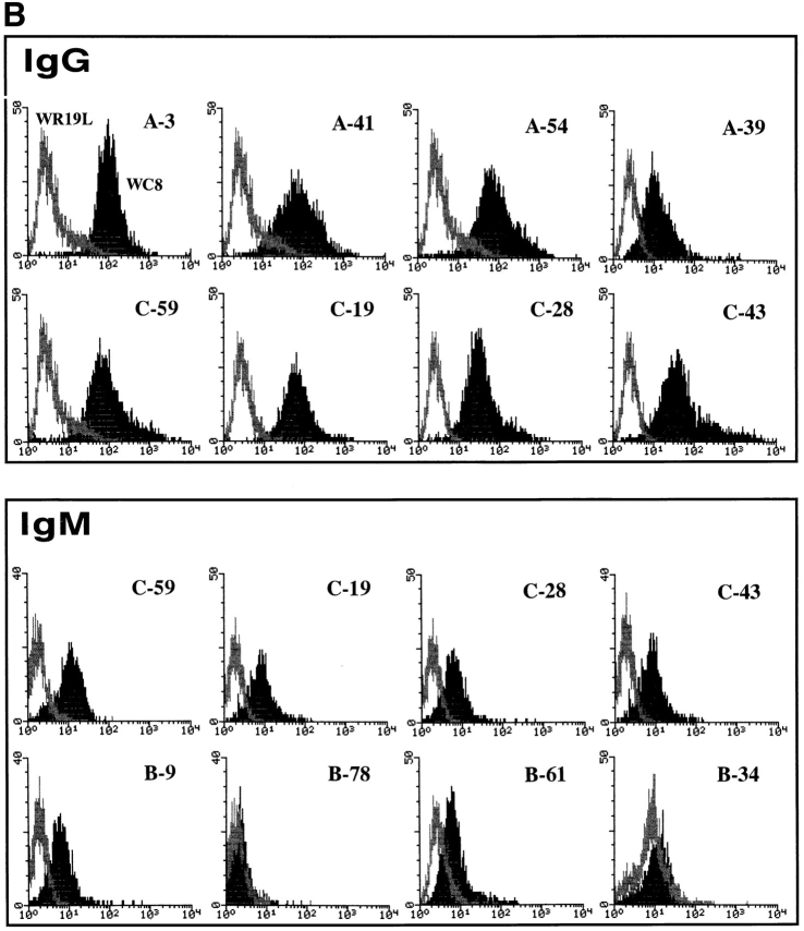
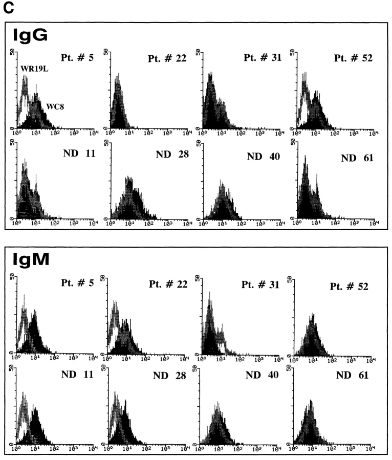
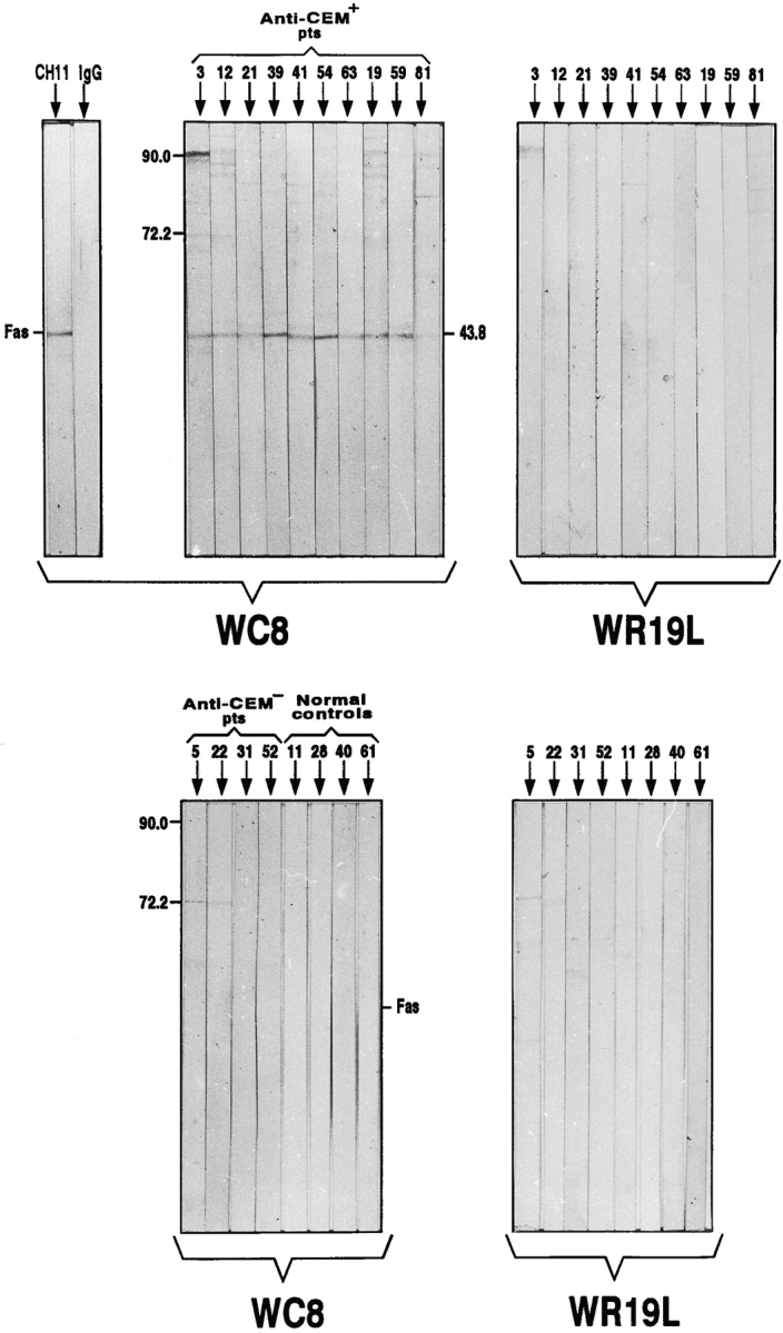
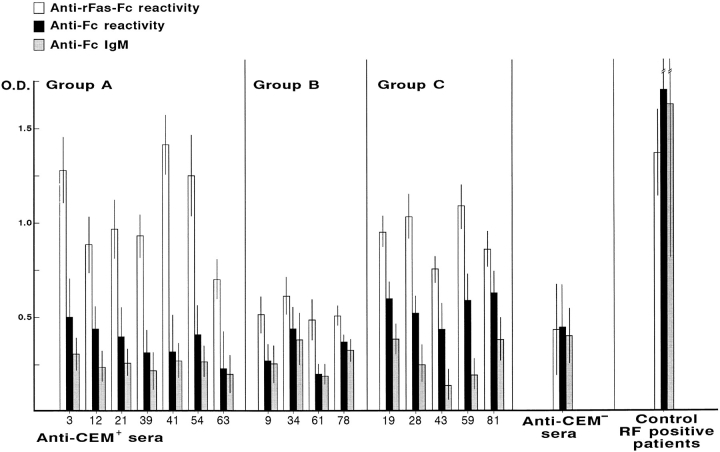
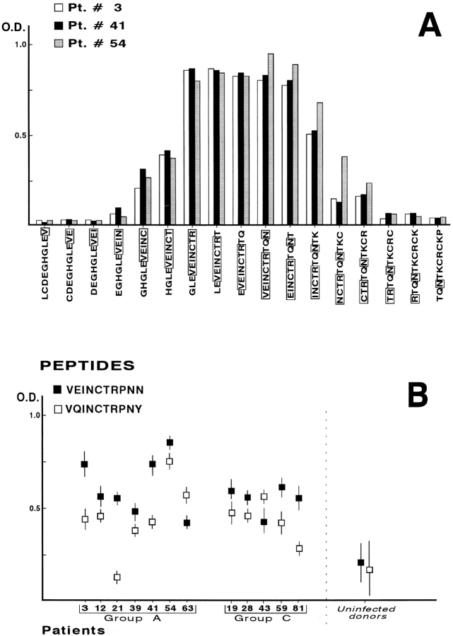
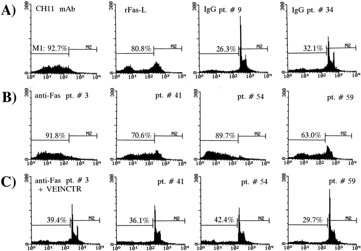
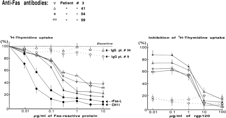
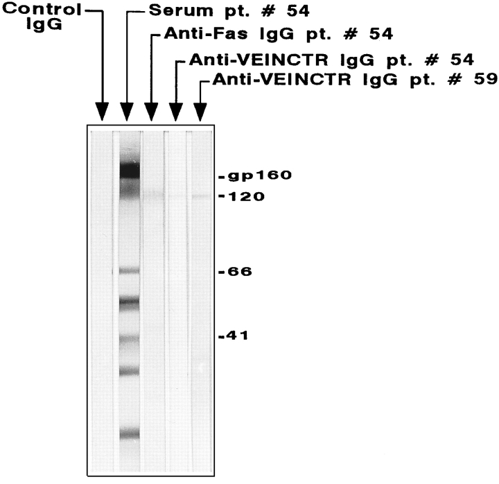
Previous studies have demonstrated that T cell-reactive antibodies in HIV-1 infection contribute to lymphocyte depletion by cytotoxicity that involves differential membrane targets, such as the 43.5-kD receptor on CEM cells. Here, we show that these antibodies bind Fas as result of a molecular mimicry of the gp120. Both flow cytometry and immunoblotting using the human Fas-transfected mouse WC8 lymphoma revealed positive binding of immunoglobulin G from several patients to a 43.8-kD membrane receptor that also reacts with the CH11 anti-Fas monoclonal antibody. Specificity to Fas was further confirmed to chimeric recombinant human Fas-Fc by ELISA, whereas overlapping peptide mapping of a Fas domain (VEINCTR-N) shared by gp120 V3 loop demonstrated a predominant affinity to the full-length 10-mer peptide. Four anti-Fas affinity preparations greatly increased the subdiploid DNA peak of CEM cells similar to agonist ligands of Fas. In addition, anti-Fas immunoglobulin G strongly inhibited the [3H]thymidine uptake of CEM cells in proliferative assays, inducing a suppression as high as provoked by both CH11 mAb and recombinant human Fas ligand. Since anti-Fas were reactive to gp120, it is conceivable that antibodies binding that domain within the V3 region are effective cross-linkers of Fas and increase apoptosis in peripheral T cells. These results suggest that autologous stimulation of the Fas pathway, rather than of lymphocytotoxic antibodies, may aggravate lymphopenia in a number of HIV-1+ subjects.


Figures

Figure 1
Figure 1
Distribution of serum IgG and IgM reactivities by ELISA to CEM cell membrane in 74 HIV-1–infected individuals at different CDC stages. Positive antibody titers were observed in 16 patients divided into groups A, B, and C in relation to serological elevations of IgG, IgM, or both. Numbers refer to both HIV-1+ patients and two normal donors (ND) showing weak yet definite IgM elevations to the membrane. The reactivities of the remaining 58 HIV-1+ sera are included in the shaded area whose positivity limits are the mean + 3 SD of OD values obtained from 67 uninfected controls. SD of single samples are related to outlayer values.
Figure 2
Figure 2
(A) Indirect immunofluorescence assay using as substrate the human Fas transfected mouse WC8 lymphoma (top) and its Fas-negative mutant WR19L (bottom). Representative fluorescence pattern from patient 41, whose serum IgG were reactive to the WC8 membrane with no evidence of specificity to Fas-negative cells. (B) Flow cytometry analysis of Fas reactivity of T cell–reactive IgG and IgM detected by ELISA in HIV-1–infected individuals. Positive binding of IgG to the antigen occurred in virtually all patients included in groups A and C, showing high fluorescence intensity to WC8 cell, as compared to its Fas-negative parental line (WR19L). By contrast, the reactivity of IgM to WC8 was almost undetectable, since the fluorescence intensity was less than one decade of dynamic range of the Fas-negative control. (C) Cytofluorimetric control tests of patients unreactive to CEM membrane and normal donors (ND). Representative patterns from both groups consistently showed lack of IgG and IgM reactivity to the human Fas–transfected cell line (WC8). Pt., patient.
Figure 2
Figure 2
(A) Indirect immunofluorescence assay using as substrate the human Fas transfected mouse WC8 lymphoma (top) and its Fas-negative mutant WR19L (bottom). Representative fluorescence pattern from patient 41, whose serum IgG were reactive to the WC8 membrane with no evidence of specificity to Fas-negative cells. (B) Flow cytometry analysis of Fas reactivity of T cell–reactive IgG and IgM detected by ELISA in HIV-1–infected individuals. Positive binding of IgG to the antigen occurred in virtually all patients included in groups A and C, showing high fluorescence intensity to WC8 cell, as compared to its Fas-negative parental line (WR19L). By contrast, the reactivity of IgM to WC8 was almost undetectable, since the fluorescence intensity was less than one decade of dynamic range of the Fas-negative control. (C) Cytofluorimetric control tests of patients unreactive to CEM membrane and normal donors (ND). Representative patterns from both groups consistently showed lack of IgG and IgM reactivity to the human Fas–transfected cell line (WC8). Pt., patient.
Figure 3
Figure 3
Immunoblotting of anti-Fas specificities in sera from HIV-1+ patients and uninfected controls. Although in the presence of undefined bands related to weak reactivity with other antigens, the majority of IgG binding the WC8 cells in the flow cytometry assay were reactive to a 43.8-kD antigen, that was also detected by the anti-Fas mAb from clone CH11. Conversely, no evidence of reactivity was observed when using the Fas-negative membrane (WR19L) as control (upper section). Sera from patients unreactive to the CEM membrane and uninfected donors showed no evidence of binding to the transfected antigen (lower section). Numbers are related to patients and controls. IgG indicate the negative control provided by 0.5 mg/ml of polyspecific IgG incubated with the WC8-blotted membrane.
Figure 4
Figure 4
Reactivity to Fas by ELISA using the chimeric rFas-Fc protein in 16 HIV-1+ sera showing positive binding to CEM membrane as compared to 39 anti-CEM–negative sera. Specificity to Fas was predominantly expressed in all instances with respect to either total or IgM-mediated rheumatoid activity. The highest values occurred in patients from groups A (3, 41, and 54) and C (28 and 59), showing serum elevations of T cell–reactive IgG, whereas the sera from patients unreactive to CEM membrane showed mean values of weak affinity to the antigen. By contrast, control sera from four HIV-1–uninfected patients with high RF titers reacted with Fc both in its chimeric and natural forms.
Figure 5
Figure 5
(A) Epitope mapping of a domain of Fas (aa position 90–117), including an 8-mer stretch (VEINCTR—N) shared by the V3 loop of gp120. A panel of 17 overlapping 10-mer peptides used as antigens in ELISA revealed a predominant affinity to that domain in sera 3, 41, and 54 although IgG from patient 54 showed a slight increase of reactivity to a subsequent peptide constructed with (N) Asn in the 108 aa position. Specificity to the VEINCTR peptide was almost undetectable in sera from patients unreactive to the CEM membrane. (B) ELISA reactivity to synthetic peptides resembling the homology sequence of the aa 99–108 domain of Fas in the V3 loop of HIV-1iiib (VEINCTRPNN) and HIV-1mn (VQINCTRPNY) strains. OD values to both peptides were apparently equivalent in most of the anti-Fas IgG in groups A and C. In contrast, sera 3, 21, and 41 showed a slight difference in their affinity to the peptides, suggesting that the replacement of E (Glu) by Q (Gln) was influencing the antigenicity of the epitope.
Figure 6
Figure 6
SDS-PAGE pattern of isolated anti-Fas antibody preparations obtained by absorption of sera on affinity columns coupled with the Fas+ membrane lysate from WC8 cells and further absorption on WR19L lysate columns for clearance of nonspecific reactivities. A prevalent IgG content was observed in those preparations, even though traces of IgM were also present. Parallel absorptions using VEINCTR peptide–coupled affinity columns provided eluates showing a similar content of antibodies in sera 54 and 59. Standard IgM and IgG were myeloma proteins.
Figure 7
Figure 7
Functional studies of isolated anti-Fas molecules from patients 3, 41, 54, and 59. (A) Percentage of apoptotic CEM cells measured as the extent of PI subdiploid DNA staining (M1) induced by 20 μg/ml of agonist ligands of Fas (CH11 mAb and rFas-L) compared to the effect of control IgG from two patients unreactive to CEM membrane. (B) The extent of subdiploid DNA in cell preparations that were treated with similar concentrations of the purified anti-Fas from the four patients ranged from ∼63–92%, and was apparently equivalent to the effect of functional ligands. (C) Preincubation of anti-Fas preparations with 7.5 μg/ml of VEINCTR peptide removed the ability of those antibodies to increase the PI subdiploid DNA peak, suggesting their functional saturation by the related antigen.
Figure 8
Figure 8
Inhibition of CEM proliferative rate by anti-Fas preparations. Significant inhibition (P <0.02) was noted in cell suspensions that were incubated with anti-Fas IgG from patients 41 and 54 by the concentration of 1 μg/ml. A moderate but significant effect (P <0.05) was also recorded using anti-Fas preparations 3 and 59 at higher concentrations. The test included both positive and negative controls. The effect of both CH11 mAb and rFas-L was nearly equivalent to anti-Fas antibodies from patients, whereas the control Fas-unreactive IgG from patients 9 and 34 failed to induce significant variations of the baseline [3H]thymidine uptake (left panel). Preincubation of anti-Fas with rgp120 induced in all instances a dose-dependent suppression of their inhibitory property on CEM proliferation, which was measured as the percentage of [3H]thymidine incorporation. The effect of control IgG was not influenced by the recombinant glycoprotein (right panel).
Figure 9
Figure 9
Immunoblotting of isolated anti-Fas IgG from patient 54 on the HIV-1 lysate, as compared with serum pattern of positivities to the viral bands. The antibody was found to react with gp120 in a fashion similar to both antibody preparations (from patients 54 and 59) absorbed on VEINCTR columns. Negative control using commercial human IgG was also provided.

Similar articles

Cited by

References

    1. Laurent-Crawford AG, Krust B, Muller S, Riviere Y, Rey-Cuillè MA, Bechet JM, Montagnier L, Hovanessian AG. The cytopathic effect of HIV is associated with apoptosis. Virology. 1991;185:829–838. - PubMed
    1. Terai C, Kornbluth RS, Pauza CD, Richman DD, Carson DA. Apoptosis as a mechanism of cell death in cultured T lymphoblasts acutely infected with HIV-1. J Clin Invest. 1991;87:1710–1721. - PMC - PubMed
    1. Meyaard L, Otto SA, Jonker RR, Mijnster MJ, Keet RP, Miedema F. Programmed death of T cells in HIV-1 infection. Science (Wash DC) 1992;257:217–219. - PubMed
    1. Gougeon ML, Laurent-Crawford AG, Hovanessian AG, Montagnier L. Direct and indirect mechanism mediating apoptosis during HIV infection: contribution to in vivo CD4 T cell depletion. Semin Immunol. 1993;5:187–196. - PubMed
    1. Groux H, Torpier G, Monte D, Mouton Y, Capron A, Ameisen JC. Activation-induced death by apoptosis in CD4+T cells from human immunodeficiency virus– infected asymptomatic individuals. J Exp Med. 1992;175:331–340. - PMC - PubMed

Publication types

MeSH terms