. 1996 Dec 1;184(6):2311-26.
doi: 10.1084/jem.184.6.2311.

Basement membrane and repair of injury to peripheral nerve: defining a potential role for macrophages, matrix metalloproteinases, and tissue inhibitor of metalloproteinases-1

Affiliations

Basement membrane and repair of injury to peripheral nerve: defining a potential role for macrophages, matrix metalloproteinases, and tissue inhibitor of metalloproteinases-1

M La Fleur et al. J Exp Med. .

Abstract

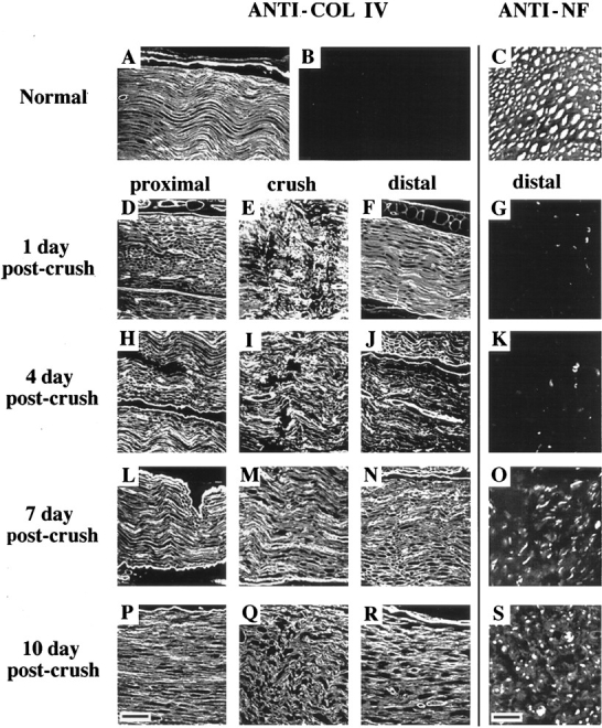
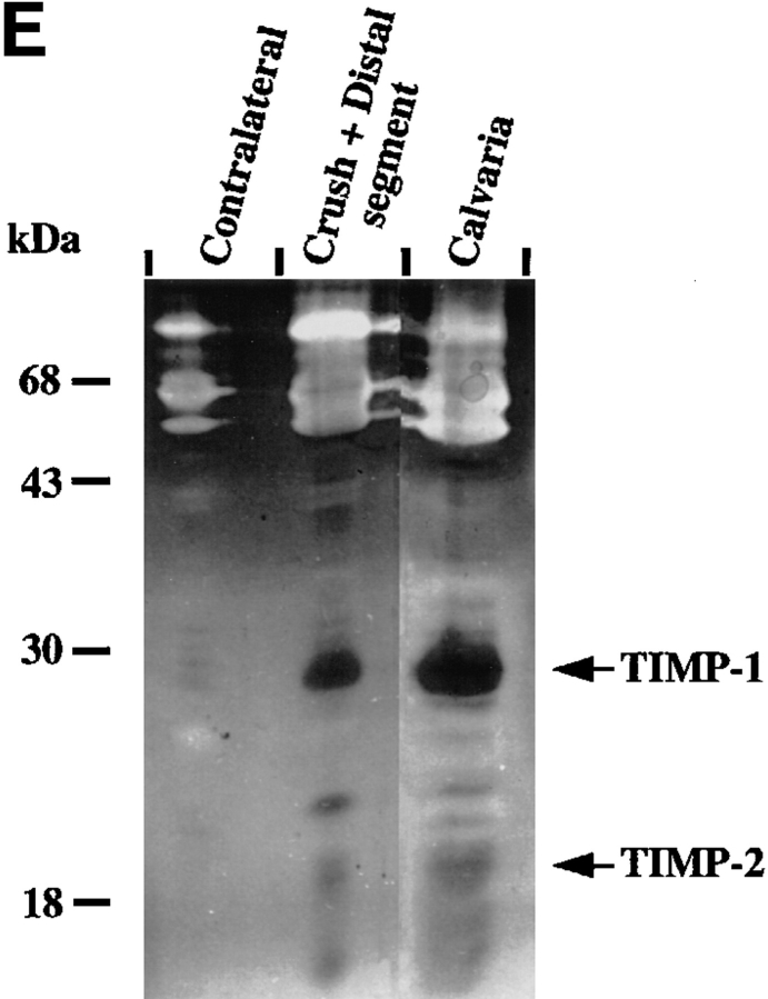
Injury to a peripheral nerve is followed by a remodeling process consisting of axonal degeneration and regeneration. It is not known how Schwann cell-derived basement membrane is preserved after injury or what role matrix metalloproteinases (MMPs) and their inhibitors play in axonal degeneration and regeneration. We showed that the MMPs gelatinase B (MMP-9), stromelysin-1 (MMP-3), and the tissue inhibitor of MMPs (TIMP)-1 were induced in crush and distal segments of mouse sciatic nerve after injury. TIMP-1 inhibitor activity was present in excess of proteinase activity in extracts of injured nerve. TIMP-1 protected basement membrane type IV collagen from degradation by exogenous gelatinase B in cryostat sections of nerve in vitro. In vivo, during the early phase (1 d after crush) and later phase (4 d after crush) after injury, induction of TNF-alpha and TGF-beta 1 mRNAs, known modulators of TIMP-1 expression, were paralleled by an upregulation of TIMP-1 and gelatinase B mRNAs. At 4 days after injury, TIMP-1, gelatinase B, and TNF-alpha mRNAs were localized to infiltrating macrophages and Schwann cells in the regions of nerve infiltrated by elicited macrophages. TIMP-1 and cytokine mRNA expression was upregulated in undamaged nerve explants incubated with medium conditioned by macrophages or containing the cytokines TGF-beta 1, TNF-alpha, and IL-1 alpha. These results show that TIMP-1 may protect basement membrane from uncontrolled degradation after injury and that cytokines produced by macrophages may participate in the regulation of TIMP-1 levels during nerve repair.


Figures

Figure 1
Figure 1
BM integrity and axonal regeneration after sciatic nerve injury. For visualization of BM, longitudinal paraffin sections of uninjured contralateral nerve (A) and injured nerve (proximal, crush, and distal) at 1, 4, 7, and 10 days post-crush were stained with anti-COL IV antibody (D–F, H–J, L–N, and P–R, respectively). (B) A preimmune IgG control. Bar (P), 45 μm. For visualization of axons, transverse paraffin sections of uninjured contralateral nerve (C) and injured nerve approximately 5 mm distal to the crush site at 1, 4, 7, and 10 d after crush (G, K, O, and S, respectively), were stained with anti-neurofilament (ANTI-NF) antibody and visualized by immunofluorescence. Bar (S), 10 μm.
Figure 2
Figure 2
MMPs and TIMPs in normal and injured sciatic nerve. (A) Uninjured contralateral nerve, sham-operated nerve, and injured nerve at 1 and 4 d post-crush were dissected and cut into segments as described in Materials and Methods. Tissue extracts (20 μg/ lane) prepared from segments of contralateral nerve and proximal-crush-distal segments of injured nerve were assayed for gelatin-degrading activity by SDS–substrate gel zymography. Samples were treated with (+) or without (−) APMA for 1 h to partially activate latent MMP activity before electrophoresis. After electrophoresis, the gel was cut, and a portion of the gel was incubated either with (+) or without (−) 50 μM GM6001, an inhibitor of MMP activity. Clear (white) bands indicate proteolytic activity. Migration of gelatinase A (gel A) and gelatinase B (gel B) is indicated on the right, and molecular weight markers are indicated on the left. White arrows next to gelatinolytic bands in the crush segment indicate, from top to bottom: gelatinase B aggregates, 135-kD gelatinolytic band, progelatinase B, active gelatinase B, and progelatinase A. (B, top) Total RNA (100 ng) from the distal segment of injured nerve at 4 d after crush (open squares) or contralateral uninjured nerve (filled circles) was reverse transcribed, and equal amounts of cDNA were amplified by PCR. Semi-quantitative RT-PCR analysis of gelatinase B was performed by sequentially removing aliquots of the reaction mix after various numbers of cycles for each sample. For determination of the difference in transcript levels, two points (corresponding to equal amounts of input RNA or cDNA) within the exponential range of the curve were compared. (B, bottom) An example of the ethidium bromide–stained bands. (C) Segments of uninjured contralateral and 4-d postcrush nerve were cultured for 24 h, and the serum-free CM was separated on a nondenaturing SDS–polyacrylamide gel, transferred to membranes, and analyzed by immunoblotting with an anti–stromelysin-1 antibody. Prostromelysin-1 was detected in unconcentrated medium for all samples. As a positive control to show migration of prostromelysin-1 and active stromelysin-1, CM collected from a 24-h culture of mouse calvaria was incubated with APMA for 1 h. Equivalent volumes of CM per milligram of wet weight were loaded per lane. Molecular weight markers are indicated on the left. These experiments were performed on 2–4 nerves. (D) Degradation of 14C-labeled gelatin in solution was used to show the presence of endogenous MMP inhibitors in nerve extracts from pooled crush and distal segments of injured nerve. Various amounts of nerve extract (filled squares, contralateral; filled circles, crush) or recombinant human TIMP-1 (open circles) were incubated with 100 nM purified gelatinase B, followed by the addition of 14C-labeled gelatin substrate. After incubation, solubilized 14C-labeled products were determined. Results represent the mean ± range of two experiments. CM from calvaria (filled triangles), a rich source of TIMP-1, was used as a positive control. (E) Segments of uninjured contralateral and 4-d postcrush nerve were cultured for 24 h, and MMP inhibitory activity secreted in the serum-free CM was assayed by reverse zymography. CM collected from a 24 h culture of mouse calvaria served as a control. CM was concentrated 50-fold by quinine sulfate precipitation. Equivalent volumes of CM per milligram wet weight of nerve were loaded per lane. Clear (white) areas indicate proteolytic activity, and dark areas indicate MMP inhibitory activity. Molecular weight standards are indicated on the left and migration of TIMP-1 and TIMP-2 standards on the right. (F) Expression of TIMP-1 and gelatinase B mRNA in sciatic nerve at 1 and 4 d after crush. Total RNA (10 μg) from segments of uninjured contralateral nerve and proximal, crush, and distal segments of injured nerve at 1 and 4 d after crush was prepared for RNA blot analysis. RNA isolated from contalateral nerve at 1 and 4 d after crush was pooled, as was the RNA from the proximal segment of injured nerve. (Upper panel) The blot was hybridized with the following cDNA probes: TIMP-1, gelatinase B, and 28 S RNA. The blot was exposed 7 d for TIMP-1 and 10 d for gelatinase B. (Lower panel) Quantification of the mRNA signals shown was obtained by scanning of the probed blots in a PhosphorImager. The values obtained for TIMP-1 and gelatinase B in contralateral nerve was set equal to 1. Values were normalized against the value obtained for the 28 S RNA hybridization to correct for differences in loading of the different RNA samples, and are shown as fold induction, which is the ratio of mRNA in crush and distal segments of injured nerve to that of the contralateral nerve.
Figure 2
Figure 2
MMPs and TIMPs in normal and injured sciatic nerve. (A) Uninjured contralateral nerve, sham-operated nerve, and injured nerve at 1 and 4 d post-crush were dissected and cut into segments as described in Materials and Methods. Tissue extracts (20 μg/ lane) prepared from segments of contralateral nerve and proximal-crush-distal segments of injured nerve were assayed for gelatin-degrading activity by SDS–substrate gel zymography. Samples were treated with (+) or without (−) APMA for 1 h to partially activate latent MMP activity before electrophoresis. After electrophoresis, the gel was cut, and a portion of the gel was incubated either with (+) or without (−) 50 μM GM6001, an inhibitor of MMP activity. Clear (white) bands indicate proteolytic activity. Migration of gelatinase A (gel A) and gelatinase B (gel B) is indicated on the right, and molecular weight markers are indicated on the left. White arrows next to gelatinolytic bands in the crush segment indicate, from top to bottom: gelatinase B aggregates, 135-kD gelatinolytic band, progelatinase B, active gelatinase B, and progelatinase A. (B, top) Total RNA (100 ng) from the distal segment of injured nerve at 4 d after crush (open squares) or contralateral uninjured nerve (filled circles) was reverse transcribed, and equal amounts of cDNA were amplified by PCR. Semi-quantitative RT-PCR analysis of gelatinase B was performed by sequentially removing aliquots of the reaction mix after various numbers of cycles for each sample. For determination of the difference in transcript levels, two points (corresponding to equal amounts of input RNA or cDNA) within the exponential range of the curve were compared. (B, bottom) An example of the ethidium bromide–stained bands. (C) Segments of uninjured contralateral and 4-d postcrush nerve were cultured for 24 h, and the serum-free CM was separated on a nondenaturing SDS–polyacrylamide gel, transferred to membranes, and analyzed by immunoblotting with an anti–stromelysin-1 antibody. Prostromelysin-1 was detected in unconcentrated medium for all samples. As a positive control to show migration of prostromelysin-1 and active stromelysin-1, CM collected from a 24-h culture of mouse calvaria was incubated with APMA for 1 h. Equivalent volumes of CM per milligram of wet weight were loaded per lane. Molecular weight markers are indicated on the left. These experiments were performed on 2–4 nerves. (D) Degradation of 14C-labeled gelatin in solution was used to show the presence of endogenous MMP inhibitors in nerve extracts from pooled crush and distal segments of injured nerve. Various amounts of nerve extract (filled squares, contralateral; filled circles, crush) or recombinant human TIMP-1 (open circles) were incubated with 100 nM purified gelatinase B, followed by the addition of 14C-labeled gelatin substrate. After incubation, solubilized 14C-labeled products were determined. Results represent the mean ± range of two experiments. CM from calvaria (filled triangles), a rich source of TIMP-1, was used as a positive control. (E) Segments of uninjured contralateral and 4-d postcrush nerve were cultured for 24 h, and MMP inhibitory activity secreted in the serum-free CM was assayed by reverse zymography. CM collected from a 24 h culture of mouse calvaria served as a control. CM was concentrated 50-fold by quinine sulfate precipitation. Equivalent volumes of CM per milligram wet weight of nerve were loaded per lane. Clear (white) areas indicate proteolytic activity, and dark areas indicate MMP inhibitory activity. Molecular weight standards are indicated on the left and migration of TIMP-1 and TIMP-2 standards on the right. (F) Expression of TIMP-1 and gelatinase B mRNA in sciatic nerve at 1 and 4 d after crush. Total RNA (10 μg) from segments of uninjured contralateral nerve and proximal, crush, and distal segments of injured nerve at 1 and 4 d after crush was prepared for RNA blot analysis. RNA isolated from contalateral nerve at 1 and 4 d after crush was pooled, as was the RNA from the proximal segment of injured nerve. (Upper panel) The blot was hybridized with the following cDNA probes: TIMP-1, gelatinase B, and 28 S RNA. The blot was exposed 7 d for TIMP-1 and 10 d for gelatinase B. (Lower panel) Quantification of the mRNA signals shown was obtained by scanning of the probed blots in a PhosphorImager. The values obtained for TIMP-1 and gelatinase B in contralateral nerve was set equal to 1. Values were normalized against the value obtained for the 28 S RNA hybridization to correct for differences in loading of the different RNA samples, and are shown as fold induction, which is the ratio of mRNA in crush and distal segments of injured nerve to that of the contralateral nerve.
Figure 3
Figure 3
TIMP-1 protection of nerve BM from degradation by gelatinase B. Unfixed cryosections of uninjured sciatic nerve were incubated overnight in medium and 1 mM APMA without gelatinase B (A and B), with 0.5 nM gelatinase B (C and D), or with 0.5 nM gelatinase B and 0.5 nM recombinant TIMP-1 (E and F). Sections were then stained with anti–laminin (LN) antibody (A, C, and E) or anti-COL IV antibody (B, D, and F) and visualized by immunofluorescence. Bars in A (A and B) 50 μm; (for C–F) 20 μm.
Figure 4
Figure 4
Kinetic analysis of the expression of TIMP-1, gelatinase B, and other injury-related genes by means of semi-quantitative PCR. mRNA transcripts for ApoE, c-fms, GAPDH, gelatinase B, TIMP-1, TNF-α, TGF-β1, and NGF-β were identified in the crush and distal segments of injured sciatic nerve relative to the contralateral nerve by semiquantitative PCR as described in the legend to Fig. 2 B. Each point represents the mean of three independent experiments. Bars, ± SEM.
Figure 5
Figure 5
Expression of TIMP-1 mRNA and localization of macrophages to injured sciatic nerve. (A) Longitudinal cryosection of 4-d postcrush nerve was hybridized with digoxigenin-labeled antisense RNA probe to TIMP-1. The proximal, crush, and distal segments of the injured nerve are indicated. The bracket indicates the crush site. The arrow points to the India ink at the crush site. TIMP-1 expression is seen at the crush site and in the distal segment of injured nerve. The distal segment of the section has a fragmented appearance and loss of some tissue because of poor adherence of nerve sections to the slide. (B) Immunohistochemical staining of macrophages in sections of injured nerve by means of a macrophage-specific antibody F4/80, and a horseradish peroxidase–labeled secondary antibody. Increased numbers of macrophages are seen at the crush site and in the distal segment. Bar, 100 μm.
Figure 6
Figure 6
Localization of TIMP-1, gelatinase B, and TNF-α mRNA in the distal segment of injured nerve by in situ hybridization. Serial longitudinal cryosections of 4-d post-crush nerve were hybridized with digoxigenin-labeled antisense RNA probes to TIMP-1, gelatinase B, and TNF-α and counterstained with Hoechst 33258. (A) Hematoxylin-and-eosin staining of injured nerve. The enclosed areas in A, the crush site (*) and distal segment, are shown at higher magnification in B and D–S. D, H, L, and P, marked with * show the crush sites. E–G, I–K, M–O, and Q–S are distal segments. (B) Control showing the crush site hybridized with TIMP-1 sense probe. The black material at the top is the India ink marking the injury site. (C) Section of contralateral nerve hybridized with TIMP-1 antisense probe. (D) Crush and (E) distal segments hybridized with antisense TIMP-1 probe. (F) Hybridization with gelatinase B and (G) TNF-α antisense probes in the distal segment. (H–K) Hoechst nuclear staining (fluorescence) of sections in (D–G). (L–O) Corresponding staining for macrophages with mAb F4/80 on adjacent sections. (P–S) Corresponding staining for Schwann cells with S-100 antibody on adjacent sections. Thick arrows indicate cells expressing mRNA, and thin arrows indicate nonexpressing cells. Note that all macrophages express TIMP-1, gelatinase B, or TNF-α. Bars (A) 200 μm; (P) (for B–S), 15 μm.

References

    1. Fawcett JW, Keynes RJ. Peripheral nerve regeneration. Annu Rev Neurosci. 1990;13:43–60. - PubMed
    1. Griffin, J.W., and D.N. Hoffman. 1993. Degeneration and regeneration in the peripheral nervous system. In Peripheral Neuropathy. 3rd ed. P.J. Dyck, P.K. Thomas, J.W. Griffin, P.A. Low, and J.F. Poduslo, editors. W.B. Saunders, Philadelphia. 361–376.
    1. Bunge RP. Expanding roles for the Schwann cell: ensheathment, myelination, trophism and regeneration. Curr Opin Neurobiol. 1993;3:805–809. - PubMed
    1. Sanes JR. Extracellular matrix molecules that influence neural development. Annu Rev Neurosci. 1989;12:491–516. - PubMed
    1. Martini R. Expression and functional roles of neural cell surface molecules and extracellular matrix components during development and regeneration of peripheral nerves. J Neurocytol. 1994;23:1–28. - PubMed

Publication types

MeSH terms