. 1996 Dec 1;184(6):2341-51.
doi: 10.1084/jem.184.6.2341.

The route of antigen entry determines the requirement for L-selectin during immune responses

Affiliations

The route of antigen entry determines the requirement for L-selectin during immune responses

M D Catalina et al. J Exp Med. .

Abstract

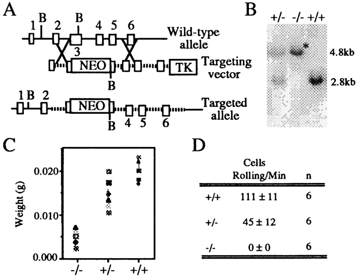
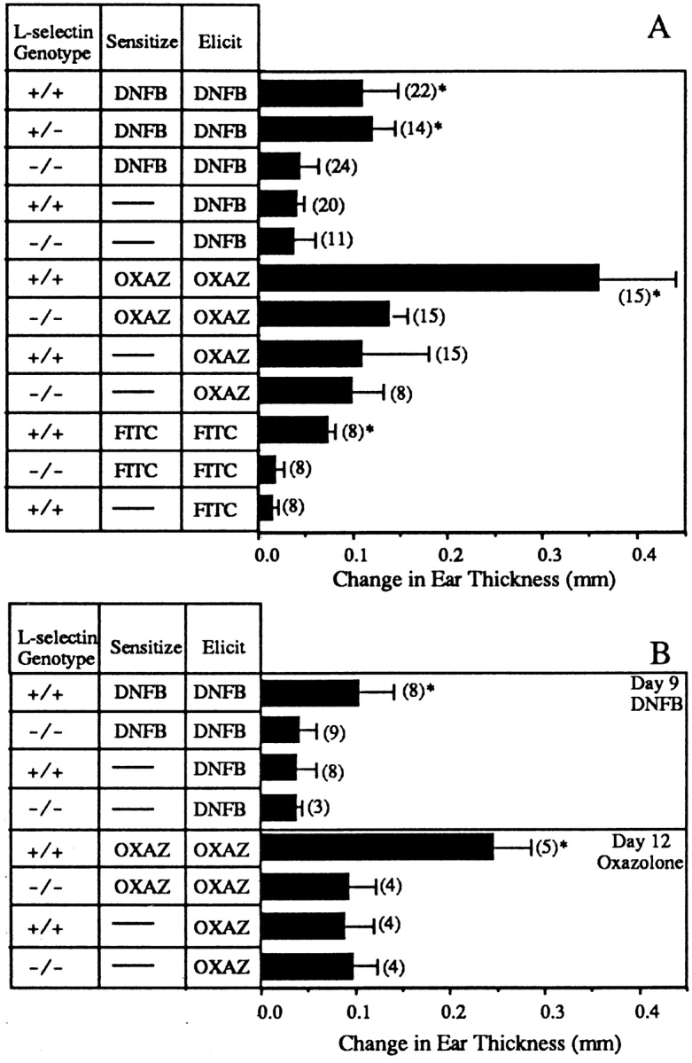
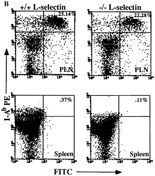
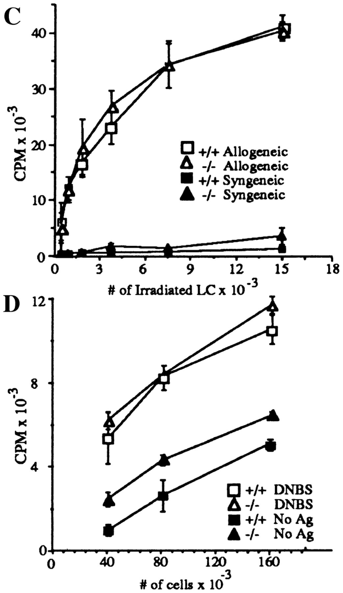
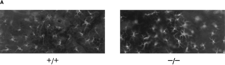
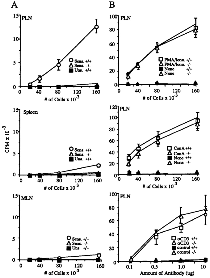
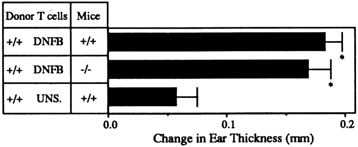
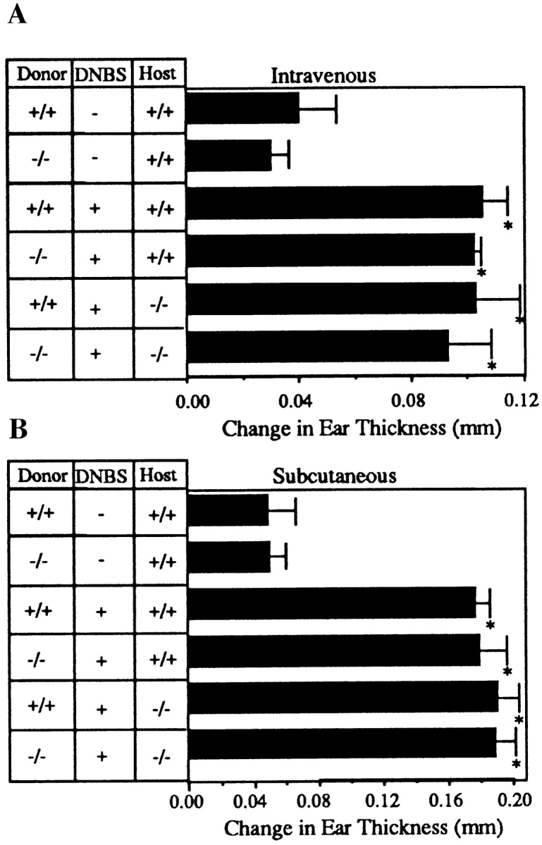
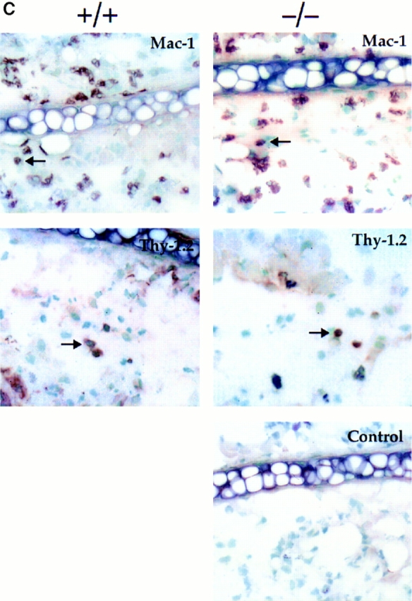
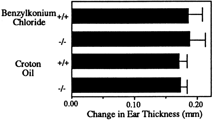
L-selectin, an adhesion molecule constitutively expressed on leukocytes, is important for primary adhesion and extravasation of lymphocytes at specialized high endothelial venules within lymph nodes and other leukocytes at sites of inflammation. We have generated L-selectin-deficient mice by targeted disruption, and have confirmed a previously reported phenotype which includes strikingly impaired contact hypersensitivity (CHS) responses to reactive haptens (Tedder, T.F., D.A. Steeber, and P. Pizcueta. 1995. J. Exp. Med. 181:2259-2264; Xu, J.C., I.S. Grewal, G.P. Geba, and R.A. Flavell. 1996. 183:589-598.). Since the mechanism of this impairment has not been clarified, we sought to define the stage(s) at which the CHS response is affected in L-selectin-deficient mice. We show that epidermal Langerhans cells in L-selectin-deficient mice are normal in number, migrate to peripheral lymph nodes appropriately, and are functional in presenting allogeneic and haptenic antigens. Moreover, T cells, as well as neutrophil and monocyte effector populations, are fully capable of entry into the inflamed skin sites in the absence of L-selectin. Thus, antigen presentation and effector mechanisms are intact in L-selectin deficient mice. In contrast, virtually no antigen-specific T cells can be found within draining peripheral nodes after a contact challenge, suggesting that the defect resides primarily in the inability of antigen-specific T cells to home to and be activated in these nodes. Indeed, L-selectin-deficient mice mount completely normal CHS responses when alternate routes of immunization are used. These studies pinpoint the lesion in CHS to a discrete stage of the afferent limb of the response, clarify the role of L-selectin on effector populations, and illustrate the critical importance of the route of antigen entry to the successful execution of an immune response.


Figures

Figure 1
Figure 1
Generation and characterization of L-selectin deficient mice. (A)The neomycin resistance gene driven by the PGK promoter was inserted into the lectin domain, exon 3, of a targeting vector containing 6.5 kb of L-selectin genomic DNA. The HSV-thymidine kinase gene was added to the 3′ end of the replacement vector as a negative selection marker. (B) Southern hybridization of L-selectin–deficient mouse DNA shows only the mutated L-selectin allele. Mouse-tail DNA was digested with BglII and hybridized with a radiolabeled intron-1 probe. Only the mutant 4.8-kb allele is present in the homozygous L-selectin–deficient mouse (*). Both the mutant 4.8-kb and the germline 2.8-kb allele are present in the heterozygous mouse. (C ) Peripheral nodes (inguinal, brachial, and axillary) were excised from L-selectin–deficient, heterozygous, and wild-type littermate mice and weighed. Both L-selectin wild-type and heterozygous mice have significantly increased weights over L-selectin mutant animals (n = 9; P <0.05). (D) Primary adhesion of L-selectin– deficient, heterozygous mutant, and wild-type spleen cells to PNAd. Cells were analyzed under shear stress in coated glass capillary tubes at 2.1 dynes/cm2 (40). Wild-type mice have signifcantly greater numbers of splenocytes rolling on PNAd-coated surfaces than either heterozygous or homozygous L-selectin–deficient mice (P <0.05).
Figure 2
Figure 2
(A) L-selectin–deficient mice have impaired CHS responses to DNFB, oxazolone, and FITC. Wild-type mice sensitized and elicited had significantly greater ear swelling than the sensitized and elicited L-selectin–deficient mice (*; P <0.05) and significantly greater ear swelling than both wild-type and L-selectin–deficient elicited-only mice (*; P <0.05). There was no significant difference between the response of L-selectin– deficient mice which were both sensitized and elicited, and either wildtype or L-selectin–deficient mice which were elicited only for any of the tested haptens (P >0.5). Results shown are the change in ear thickness at 24 h from mice elicited with CHS reagent on days 5 (DNFB), 6 (FITC), or 7 (oxazolone). n values are given in parentheses. (B) Delay of challenge with either DNFB or oxazolone does not restore the ability of L-selectin– deficient mice to respond to CHS agents. Wild-type mice sensitized and elicited had significantly greater ear swelling than the sensitized and elicited L-selectin–deficient mice when mice were challenged 9 d after sensitization for DNFB or 12 d after sensitization for oxazolone (*; P <0.05). There was no significant difference between the response of sensitized and elicited L-selectin–deficient mice and mice which were elicited only (P >0.5).
Figure 3
Figure 3
L-selectin–deficient mice have an intact antigen presentation pathway. (A) Epidermal whole mounts from L-selectin–deficient mice show normal numbers and distribution of LC. Whole mounts of abdominal skin were prepared and stained with anti-I-Ab (41). I-A+ cells were counted in 30 fields/slide (field = 0.1 mm2), and calculated as the average of cells/mm2 over two slides. (B) LC are present in L-selectin–deficient mice and drain to PLN following skin painting with FITC. Mice were sensitized with FITC and 24 h later PLN and spleens were collected and centrifuged over metrizamide gradients (18). Cells at the interface were collected and stained with biotinylated anti– mouse I-A followed by streptavidin-PE and analyzed by twocolor FACS® analysis to detect the presence of draining I-A+/ FITC+ LC. One of six representative experiments is shown. (C ) LC from L-selectin–deficient mice can initiate allogeneic responses. LC collected from PLN of FITC-painted wild-type (squares) or L-selectin–deficient mice (triangles) were irradiated and incubated for 48 h with 2 × 10⁵ T cells from either BALB/c (allogeneic; open squares and triangles) or C57BL/6J (syngeneic; closed squares and triangles) mice. Cultures were pulsed with [3H]thymidine for 18 h before harvesting. (D) LC can initiate antigen-specific responses. Irradiated PLN cells were used as APCs from wild-type or L-selectin–deficient mice, and were either pulsed with or without DNBS. These cells were incubated for 48 h with increasing concentrations of PLN T cells isolated from wild-type mice sensitized with DNFB 5 d before. Cultures were pulsed with [3H]thymidine for 18 h before harvesting.
Figure 3
Figure 3
L-selectin–deficient mice have an intact antigen presentation pathway. (A) Epidermal whole mounts from L-selectin–deficient mice show normal numbers and distribution of LC. Whole mounts of abdominal skin were prepared and stained with anti-I-Ab (41). I-A+ cells were counted in 30 fields/slide (field = 0.1 mm2), and calculated as the average of cells/mm2 over two slides. (B) LC are present in L-selectin–deficient mice and drain to PLN following skin painting with FITC. Mice were sensitized with FITC and 24 h later PLN and spleens were collected and centrifuged over metrizamide gradients (18). Cells at the interface were collected and stained with biotinylated anti– mouse I-A followed by streptavidin-PE and analyzed by twocolor FACS® analysis to detect the presence of draining I-A+/ FITC+ LC. One of six representative experiments is shown. (C ) LC from L-selectin–deficient mice can initiate allogeneic responses. LC collected from PLN of FITC-painted wild-type (squares) or L-selectin–deficient mice (triangles) were irradiated and incubated for 48 h with 2 × 10⁵ T cells from either BALB/c (allogeneic; open squares and triangles) or C57BL/6J (syngeneic; closed squares and triangles) mice. Cultures were pulsed with [3H]thymidine for 18 h before harvesting. (D) LC can initiate antigen-specific responses. Irradiated PLN cells were used as APCs from wild-type or L-selectin–deficient mice, and were either pulsed with or without DNBS. These cells were incubated for 48 h with increasing concentrations of PLN T cells isolated from wild-type mice sensitized with DNFB 5 d before. Cultures were pulsed with [3H]thymidine for 18 h before harvesting.
Figure 4
Figure 4
L-selectin–deficient mice have markedly diminished sensitized T cells after skin painting with DNFB. (A) Cells from mice sensitized 5 d earlier with DNFB were cultured for 36 h in the presence of DNBS after which cultures were pulsed with [3H]thymidine for 18 h. Each point represents the average of three separate mouse groups, each group consisting of pooled organs from two mice. Each group was done in triplicate. Background proliferation, detected by incubation of the cells in the absence of DNBS, was subtracted. (B) Resident T cells from L-selectin–deficient lymph nodes are able to respond normally to in vitro stimuli. PLN were incubated with PMA plus Ionomycin, Con A, or immobilized anti-CD3 or hamster IgG control antibody. Cells were cultured in triplicate for 36 h and then pulsed with [3H]thymidine for 18 h.
Figure 5
Figure 5
Adoptive transfer of wild-type sensitized T cells into L-selectin–deficient mice restores their ability to mount CHS responses. 2 × 10⁷ T cells from wild-type mice sensitized with DNFB were harvested and injected into either wild-type or L-selectin–deficient mice. When mice were elicited 12 h later, there was no significant difference between the 24 h ear swelling response of L-selectin–deficient mice as compared to wild-type littermate controls (P >0.5). Both had significantly greater responses than animals injected with PLN cells from unsensitized mice (*, P <0.05).
Figure 6
Figure 6
L-selectin–deficient mice develop CHS responses when sensitized via an alternative route. (A) Ear swelling responses 5 d after intravenous injection of DNBS-conjugated splenic dendritic cells and challenge with DNFB were not significantly different between L-selectin– deficient and wild-type littermate recipients irrespective of the source of donor cells. Responses were significantly different from those of mice injected with splenic dendritic cells not conjugated to DNBS (*, P <0.05). (B) Ear swelling responses 5 d after subcutaneous injection of DNBS-conjugated spleen cells and challenge with DNFB were not significantly different between L-selectin–deficient and wild-type littermate recipients (P >0.5), but were significantly different from mice injected with spleen cells not conjugated to DNBS (*, P <0.05). (C ) Immunohistochemical staining of ear sections with anti-Mac-1 and anti-Thy1.2, shows that both L-selectin–deficient mice and wild-type mice injected subcutaneously with L-selectin–deficient DNP-conjugated spleen cells and elicited with DNFB, have similar numbers of infiltrating macrophages and T cells. Arrows indicate representative stained cells. Control mice were injected subcutaneously with L-selectin–deficient spleen cells not conjugated to DNBS (×400). Cells stained brown are positive for that marker.
Figure 6
Figure 6
L-selectin–deficient mice develop CHS responses when sensitized via an alternative route. (A) Ear swelling responses 5 d after intravenous injection of DNBS-conjugated splenic dendritic cells and challenge with DNFB were not significantly different between L-selectin– deficient and wild-type littermate recipients irrespective of the source of donor cells. Responses were significantly different from those of mice injected with splenic dendritic cells not conjugated to DNBS (*, P <0.05). (B) Ear swelling responses 5 d after subcutaneous injection of DNBS-conjugated spleen cells and challenge with DNFB were not significantly different between L-selectin–deficient and wild-type littermate recipients (P >0.5), but were significantly different from mice injected with spleen cells not conjugated to DNBS (*, P <0.05). (C ) Immunohistochemical staining of ear sections with anti-Mac-1 and anti-Thy1.2, shows that both L-selectin–deficient mice and wild-type mice injected subcutaneously with L-selectin–deficient DNP-conjugated spleen cells and elicited with DNFB, have similar numbers of infiltrating macrophages and T cells. Arrows indicate representative stained cells. Control mice were injected subcutaneously with L-selectin–deficient spleen cells not conjugated to DNBS (×400). Cells stained brown are positive for that marker.
Figure 7
Figure 7
L-selectin–deficient mice have a normal response to irritants. Benzylkonium chloride or croton oil was applied to mouse ears, and 16 h later measurements indicated that the acute, nonspecific ear swelling response of L-selectin–deficient mice was not significantly different from the response of wild-type littermate controls (P >0.5).

Similar articles

Cited by

References

    1. Gowans JL, Knight EJ. The route of recirculation of lymphocytes in the rat. Philos Trans R Soc Lond Ser B Biol Sci. 1964;159:257–282. - PubMed
    1. Gallatin WM, Weissman IL, Butcher EC. A cell-surface molecule involved in organ-specific homing of lymphocytes. Nature (Lond) 1983;304:30–34. - PubMed
    1. Carlos TM, Harlan JM. Leukocyte-endothelial adhesion molecules. Blood. 1994;84:2068–2101. - PubMed
    1. Von Andrian UH, Chambers JD, McEvoy LM, Bargatze RF, Arfors KE, Butcher EC. Two-step model of leukocyte-endothelial cell interaction in inflammation: distinct roles for LECAM-1 and the leukocyte beta 2 integrins in vivo. Proc Natl Acad Sci USA. 1991;88:7538–7542. - PMC - PubMed
    1. Lawrence MB, Springer TA. Leukocytes roll on a selectin at physiologic flow rates: distinction from and prerequisite for adhesion through integrins. Cell. 1991;65:859–873. - PubMed

Publication types

MeSH terms