. 1996 Dec 1;184(6):2371-84.
doi: 10.1084/jem.184.6.2371.

Viral infection of transgenic mice expressing a viral protein in oligodendrocytes leads to chronic central nervous system autoimmune disease

Affiliations

Viral infection of transgenic mice expressing a viral protein in oligodendrocytes leads to chronic central nervous system autoimmune disease

C F Evans et al. J Exp Med. .

Abstract

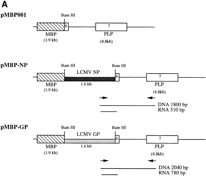
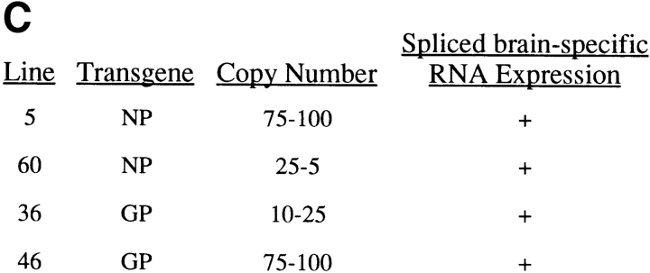
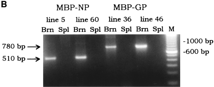
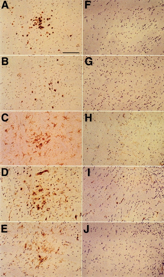
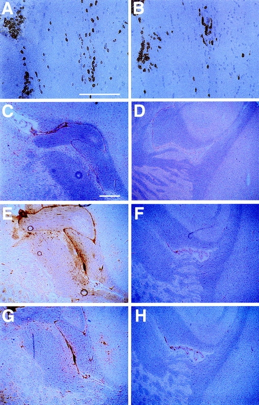
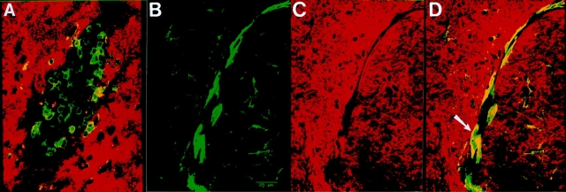
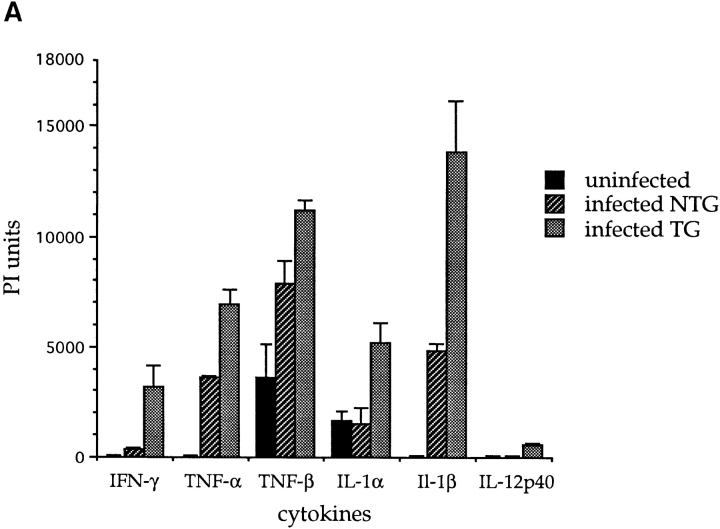
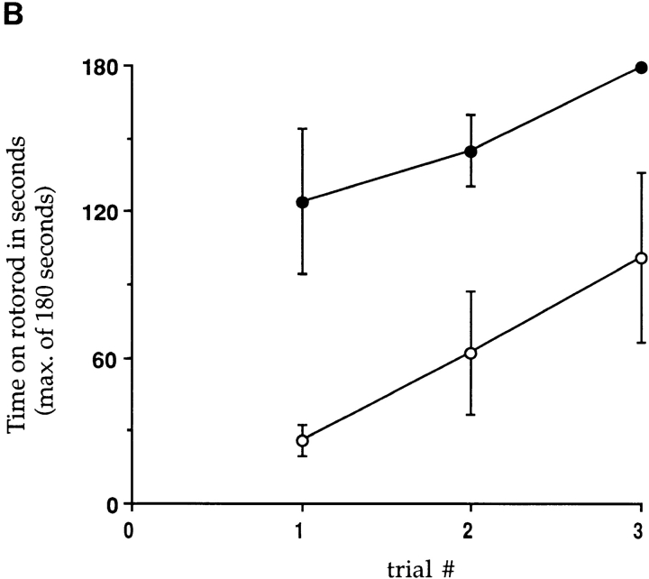
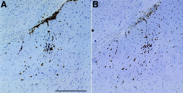
One hypothesis for the etiology of central nervous system (CNS) autoimmune disease is that infection by a virus sharing antigenic epitopes with CNS antigens (molecular mimicry) elicits a virus-specific immune response that also recognizes self-epitopes. To address this hypothesis, transgenic mice were generated that express the nucleoprotein or glycoprotein of lymphocytic choriomeningitis virus (LCMV) as self in oligodendrocytes. Intraperitoneal infection with LCMV strain Armstrong led to infection of tissues in the periphery but not the CNS, and the virus was cleared within 7-14 d. After clearance, a chronic inflammation of the CNS resulted, accompanied by upregulation of CNS expression of MHC class I and II molecules. A second LCMV infection led to enhanced CNS pathology, characterized by loss of myelin and clinical motor dysfunction. Disease enhancement also occurred after a second infection with unrelated viruses that cross-activated LCMV-specific memory T cells. These findings indicate that chronic CNS autoimmune disease may be induced by infection with a virus sharing epitopes with a protein expressed in oligodendrocytes and this disease may be enhanced by a second infection with the same or an unrelated virus. These results may explain the association of several different viruses with some human autoimmune diseases.


Figures

Figure 1
Figure 1
Summary of MBP–NP and –GP transgenic mouse lines. (A) pMBP001 contains 1.9 kb of the MBP gene promoter/enhancer sequences, and polyadenylation and splice signals from the PLP gene. The LCMV NP and GP genes were cloned into the BamHI site of pMBP001 to give pMBP–NP and pMBP–GP. Expression of spliced mRNA in transgenic mice was determined by RT-PCR using primers that hybridized within the transgene and the PLP exon 7 as indicated by arrows, with the resulting RT-PCR products of 510 bp (MBP–NP) or 780 bp (MBP–GP). (B) RNA from the brains (Brn) and spleens (Spl ) of MBP– NP and –GP transgenic mice was subjected to RT-PCR as described in Materials and Methods to detect transgene-specific spliced RNA (see A). M, molecular weight markers. (C) The transgenic mouse lines studied are listed with the relative copy number of the transgene and the results of RT-PCR transgene mRNA studies.
Figure 1
Figure 1
Summary of MBP–NP and –GP transgenic mouse lines. (A) pMBP001 contains 1.9 kb of the MBP gene promoter/enhancer sequences, and polyadenylation and splice signals from the PLP gene. The LCMV NP and GP genes were cloned into the BamHI site of pMBP001 to give pMBP–NP and pMBP–GP. Expression of spliced mRNA in transgenic mice was determined by RT-PCR using primers that hybridized within the transgene and the PLP exon 7 as indicated by arrows, with the resulting RT-PCR products of 510 bp (MBP–NP) or 780 bp (MBP–GP). (B) RNA from the brains (Brn) and spleens (Spl ) of MBP– NP and –GP transgenic mice was subjected to RT-PCR as described in Materials and Methods to detect transgene-specific spliced RNA (see A). M, molecular weight markers. (C) The transgenic mouse lines studied are listed with the relative copy number of the transgene and the results of RT-PCR transgene mRNA studies.
Figure 2
Figure 2
Immunohistochemical analyses of the brain stems of LCMV-infected MBP–NP transgenic positive and negative mice. At 12 wk after infection with LCMV, sagittal sections of the brain stem from an MBP–NP transgenic positive mouse (A–E ) and a transgenic negative mouse (F–J ) were stained with antibodies to CD8 (A and F ), CD4 (B and G ), F4/80 (C and H ), MHC class I (D and I ), and MHC class II (E and J ) as described in Materials and Methods. Bar, 100 μm; original magnification, ×200.
Figure 3
Figure 3
Immunohistochemical analyses of the brains of LCMV- infected MBP–NP mice reinfected with LCMV 6 wk after the primary infection. MBP–NP transgenic positive and negative mice were infected with LCMV; 6 wk later they were given a second LCMV infection, and then sacrificed 4 wk after second infection. Sagittal brain sections from transgenic positive (A, B, C, E, and G) and transgenic negative (D, F, and H) mice were stained with antibodies to CD8 (A), CD4 (B), F4/ 80 (C and D), MHC class I (E and F ), and MHC class II (G and H ) as described in Materials and Methods. (A and B) consecutive sections showing high power views of infiltrating lymphocytes in white matter within the internal capsule. Bar, 125 μm; original magnification, ×200. (C–H ) Low power views of the brain including the internal capsule and fimbria hippocampus. Bar, 375 μm; original magnification, ×40.
Figure 4
Figure 4
Characterization of T cell lesions by confocal microscopic analysis. An MBP–NP transgenic positive mouse was infected with LCMV, reinfected with LCMV at 6 wk after infection, and killed 12 wk after second infection. Cryocut brain sections were doubled immunofluorescently stained and analyzed by laser scanning confocal microscopy as described in Materials and Methods. (A) shows double staining of T cell lesions in the cortex with antibodies to CD8+ T cells ( green) and MBP (red ). (B, C,and D) show a region of the cortex stained with antibodies to F4/80 (B, green), MBP (C, red ), and the merged view with both labels (D, areas of colocalization of MBP and F4/80 are presented in yellow [arrow]). Bar, 25 μm.
Figure 5
Figure 5
Expression of cytokines and measurement of clinical motor dysfunction in transgenic mice infected twice with LCMV. (A) mRNAs for various cytokines were detected by RNase protection assays using RNA isolated from mouse brains as described in Materials and Methods. Nontransgenic (NTG) or transgenic (TG) mice were infected with LCMV, 6 wk later given a second LCMV infection, and sacrificed 4 wk later (n = 2–4). PI units, phosphoroimage units as defined in Materials and Methods. Values are plotted as mean ± SE. (B) MBP–NP mice were infected with LCMV and 6 wk later reinfected with LCMV. Four transgenic positive (open circles) and negative (closed circles) mice were tested for motor dysfunction on a rotorod apparatus in three consecutive trials as described in Materials and Methods. The time on the rotorod was measured, with a maximum of 180 s. The standard error is shown for all trials except the transgenic negative third trial, in which 3 out of 4 mice remained on the rod for the maximum time allowed.
Figure 5
Figure 5
Expression of cytokines and measurement of clinical motor dysfunction in transgenic mice infected twice with LCMV. (A) mRNAs for various cytokines were detected by RNase protection assays using RNA isolated from mouse brains as described in Materials and Methods. Nontransgenic (NTG) or transgenic (TG) mice were infected with LCMV, 6 wk later given a second LCMV infection, and sacrificed 4 wk later (n = 2–4). PI units, phosphoroimage units as defined in Materials and Methods. Values are plotted as mean ± SE. (B) MBP–NP mice were infected with LCMV and 6 wk later reinfected with LCMV. Four transgenic positive (open circles) and negative (closed circles) mice were tested for motor dysfunction on a rotorod apparatus in three consecutive trials as described in Materials and Methods. The time on the rotorod was measured, with a maximum of 180 s. The standard error is shown for all trials except the transgenic negative third trial, in which 3 out of 4 mice remained on the rod for the maximum time allowed.
Figure 6
Figure 6
Stimulation of increased CNS immune infiltration and activation of LCMV-memory CTL by infection with a second virus not encoding LCMV gene products. An MBP–NP transgenic positive mouse was infected with LCMV followed by VV infection 6 wk later. After another 6 wk, the mouse was sacrificed and increased numbers of CD8+ (A) and CD4+ (B) cells compared with those observed after a single LCMV infection were detected in the brain by immunohistochemical staining as described in Materials and Methods. Bar, 200 μm; original magnification ×100.

Similar articles

Cited by

References

    1. Jahnke U, Fischer EH, Alvord ECJ. Sequence homology between certain viral proteins and proteins related to encephalomyelitis and neuritis. Science (Wash DC) 1985;229:282–284. - PubMed
    1. Fujinami RS, Oldstone MBA. Amino acid homology between the encephalitogenic site of myelin basic protein and virus: a mechanism for autoimmunity. Science (Wash DC) 1985;230:1043–1045. - PubMed
    1. Fujinami RS. Virus-induced autoimmunity through molecular mimicry. Ann NY Acad Sci. 1988;540:210–217. - PubMed
    1. Oldstone MBA. Molecular mimicry and autoimmune disease. Cell. 1987;50:819–820. - PubMed
    1. Hall R. Molecular mimicry. Adv Parasitol. 1994;34:81–132. - PubMed

Publication types

MeSH terms