. 1996 Dec 1;184(6):2385-92.
doi: 10.1084/jem.184.6.2385.

Immunoglobulin G-mediated inflammatory responses develop normally in complement-deficient mice

Affiliations

Immunoglobulin G-mediated inflammatory responses develop normally in complement-deficient mice

D Sylvestre et al. J Exp Med. .

Abstract

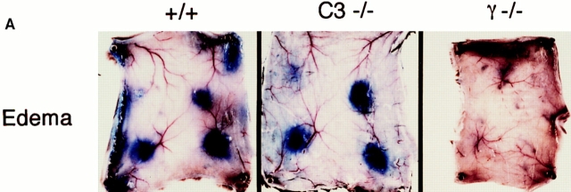
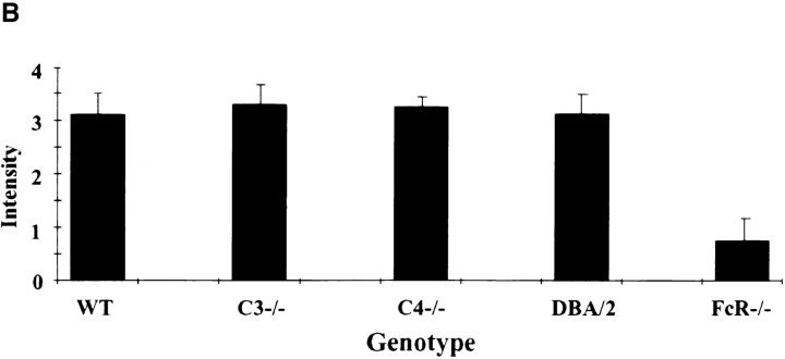
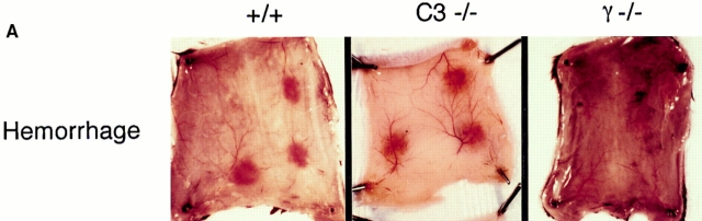
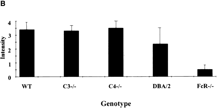
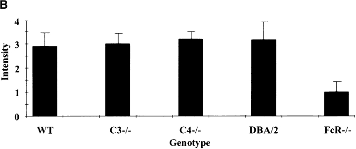
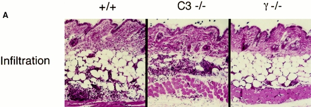
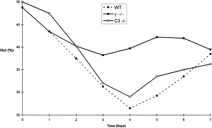
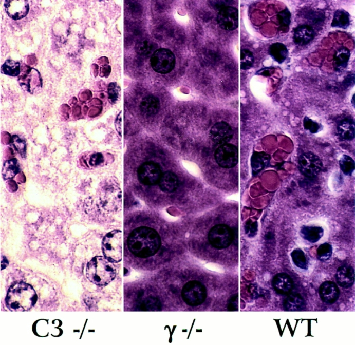
The role of complement in immunoglobulin G-triggered inflammation was studied in mice genetically deficient in complement components C3 and C4. Using the reverse passive Arthus reaction and experimental models of immune hemolytic anemia and immune thrombocytopenia, we show that these mice have types II and III inflammatory responses that are indistinguishable from those of wild-type animals. Complement-deficient and wild-type animals exhibit comparable levels of erythrophagocytosis and platelet clearance in response to cytotoxic anti-red blood cell and antiplatelet antibodies. Furthermore, in the reverse passive Arthus reaction, soluble immune complexes induce equivalent levels of hemmorhage, edema, and neutrophillic infiltration in complement-deficient and wild-type animals. In contrast, mice that are genetically deficient in the expression of Fc receptors exhibit grossly diminished reactions by both cytotoxic antibodies and soluble immune complexes. These studies provide strong evidence that the activation of cell-based Fc gamma R receptors, but not complement, are required for antibody-triggered murine inflammatory responses.


Figures

Figure 1
Figure 1
Quantitation of edema in the reverse passive Arthus reaction. (A) Macroscopic visualization for +/+ (left), complement-deficient (middle), and FcγRdeficient (right) mice. 30 mg of control rabbit IgG (upper left spot) or rabbit anti-OVA IgG was injected intradermally, followed immediately by 2 mg/kg i.v. OVA. Dormal skins were harvested after 2 h, using 1% Evan's blue in the intravenous injectate. (B) Quantitation of edema at 8 h. Microscopic sections were graded for the extent of edema on a scale of 0–4+; n >12 for each genotype, and error bars represent ± SD.
Figure 1
Figure 1
Quantitation of edema in the reverse passive Arthus reaction. (A) Macroscopic visualization for +/+ (left), complement-deficient (middle), and FcγRdeficient (right) mice. 30 mg of control rabbit IgG (upper left spot) or rabbit anti-OVA IgG was injected intradermally, followed immediately by 2 mg/kg i.v. OVA. Dormal skins were harvested after 2 h, using 1% Evan's blue in the intravenous injectate. (B) Quantitation of edema at 8 h. Microscopic sections were graded for the extent of edema on a scale of 0–4+; n >12 for each genotype, and error bars represent ± SD.
Figure 2
Figure 2
Quantitation of 8-h hemorrhage in the reverse passive Arthus reaction. (A) 30 mg of rabbit IgG (upper left spot) or rabbit anti-OVA IgG was injected intradermally, followed by 2 mg/kg i.v. OVA. Dorsal skins were harvested after 8 h. (B) Aggregated results based on the direct microscopic grading of hemorrhage on a scale of 0–4+. Results are ± SD; n >12 in each group.
Figure 2
Figure 2
Quantitation of 8-h hemorrhage in the reverse passive Arthus reaction. (A) 30 mg of rabbit IgG (upper left spot) or rabbit anti-OVA IgG was injected intradermally, followed by 2 mg/kg i.v. OVA. Dorsal skins were harvested after 8 h. (B) Aggregated results based on the direct microscopic grading of hemorrhage on a scale of 0–4+. Results are ± SD; n >12 in each group.
Figure 3
Figure 3
Quantitation of 8-h neutrophil infiltration in the reverse passive Arthus reaction. (A) 30 mg of rabbit IgG (upper left spot) or rabbit anti-OVA IgG was injected intradermally, followed by 2 mg/kg i.v. OVA. Dorsal skins were harvested after 8 h. (B) Aggregated results based on the direct microscopic grading of neutrophil infiltration on a scale of 0–4+. Results are ± SD; n >12 in each group.
Figure 3
Figure 3
Quantitation of 8-h neutrophil infiltration in the reverse passive Arthus reaction. (A) 30 mg of rabbit IgG (upper left spot) or rabbit anti-OVA IgG was injected intradermally, followed by 2 mg/kg i.v. OVA. Dorsal skins were harvested after 8 h. (B) Aggregated results based on the direct microscopic grading of neutrophil infiltration on a scale of 0–4+. Results are ± SD; n >12 in each group.
Figure 4
Figure 4
Experimental murine immune hemolytic anemia induced by rabbit polyclonal anti-MRBC IgG. Daily hematocrits of wild-type (filled squares and dotted line), homozygous γ chain–deficient (filled squares), and homozygous C3-deficient mice (open squares). Mean hematocrits obtained from five mice in each group are presented.
Figure 5
Figure 5
Histological appearance of the liver in wild-type (+/+), homozygous γ-chain–deficient (γ−/−) and homozygous C3-deficient mice (C3−/−) injected with rabbit α-MRBC IgG.
Figure 6
Figure 6
Experimental immune murine thrombocytopenia induced by mAb 6A6. Platelet counts (× 103/μl) of wild-type (filled squares and dotted line), homozygous γ chain–deficient (filled squares), and homozygous C3deficient mice (open squares). Mean data from groups of four mice are shown.

Similar articles

Cited by

References

    1. Roitt, I., J. Brostoff, and D. Male. 1993. In Immunology, 3rd ed. Mosby, Inc., London. 20.1–20.2.
    1. Snyder, E.L. 1995. Transfusion reactions. In Hematology, Basic Principles and Practice, 2nd ed. R. Hoffman, E.J. Benz, S.J. Shattol, B. Furie, H. Cohen, and L.E. Silberstein, editors. Churchill Livingstone Inc., New York.) 2045–2053.
    1. Gallin, J.I. 1993. Inflammation. In Fundamental Immunology, 3rd ed., W. Paul, editor. Raven Press, New York. 1015–1032.
    1. Arthus M. Injections repetees de serum de cheval cuez le lapin. C R Soc Biol. 1903;55:817–820.
    1. Cochrane CG, Muller-Eberhard JJ. The derivation of two distinct anaphylatoxin activities from the third and fifth components of human complement. J Exp Med. 1968;127:371–386. - PMC - PubMed

Publication types