

A role for endogenous transforming growth factor beta 1 in Langerhans cell biology: the skin of transforming growth factor beta 1 null mice is devoid of epidermal Langerhans cells
- PMID: 8976197
- PMCID: PMC2196398
- DOI: 10.1084/jem.184.6.2417
A role for endogenous transforming growth factor beta 1 in Langerhans cell biology: the skin of transforming growth factor beta 1 null mice is devoid of epidermal Langerhans cells
Abstract
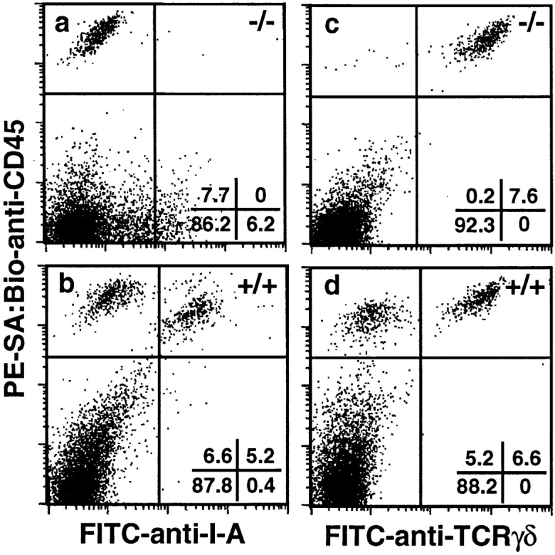
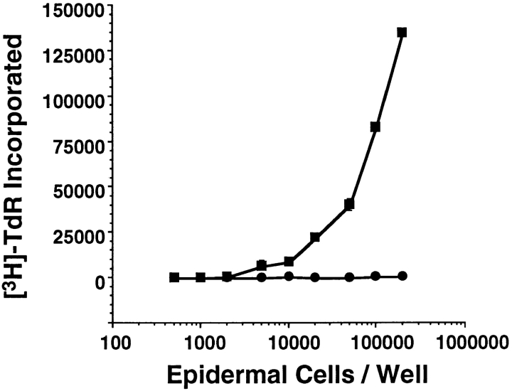
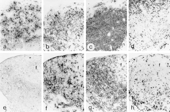
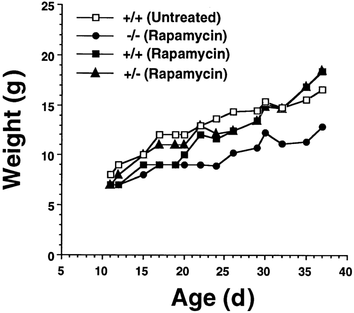
Transforming growth factor beta 1 (TGF-beta 1) regulates leukocytes and epithelial cells. To determine whether the pleiotropic effects of TGF-beta 1, a cytokine that is produced by both keratinocytes and Langerhans cells (LC), extend to epidermal leukocytes, we characterized LC (the epidermal contingent of the dendritic cell [DC] lineage) and dendritic epidermal T cells (DETC) in TGF-beta 1 null (TGF-beta 1 -/-) mice. I-A+ LC were not detected in epidermal cell suspensions or epidermal sheets prepared from TGF-beta 1 -/- mice, and epidermal cell suspensions were devoid of allostimulatory activity. In contrast, TCR-gamma delta + DETC were normal in number and appearance in TGF-beta 1 -/- mice and, importantly, DETC represented the only leukocytes in the epidermis. Immunolocalization studies revealed CD11c+ DC in lymph nodes from TGF-beta 1 -/- mice, although gp40+ DC were absent. Treatment of TGF-beta 1 -/- mice with rapamycin abrogated the characteristic inflammatory wasting syndrome and prolonged survival indefinitely, but did not result in population of the epidermis with LC. Thus, the LC abnormality in TGF-beta 1 -/- mice is not a consequence of inflammation in skin or other organs, and LC development is not simply delayed in these animals. We conclude that endogenous TGF-beta 1 is essential for normal murine LC development or epidermal localization.
Figures
References
-
- Kingsley DM. The TGF-β superfamily: new members, new receptors, and new genetic tests of function in different organisms. Genes & Dev. 1994;8:133–146. - PubMed
-
- Dickson MC, Martin JS, Cousins FM, Kulkarni AB, Karlsson S, Akhurst RJ. Defective haematopoiesis and vasculogenesis in transforming growth factor-β1 knock out mice. Development. 1995;121:1845–1854. - PubMed
Publication types
MeSH terms
Substances
Grants and funding
LinkOut - more resources
Full Text Sources
Other Literature Sources
Molecular Biology Databases
Research Materials
