. 1996 Dec 24;93(26):15069-74.
doi: 10.1073/pnas.93.26.15069.

Peroxynitrite, the coupling product of nitric oxide and superoxide, activates prostaglandin biosynthesis

Affiliations

Peroxynitrite, the coupling product of nitric oxide and superoxide, activates prostaglandin biosynthesis

L M Landino et al. Proc Natl Acad Sci U S A. .

Abstract

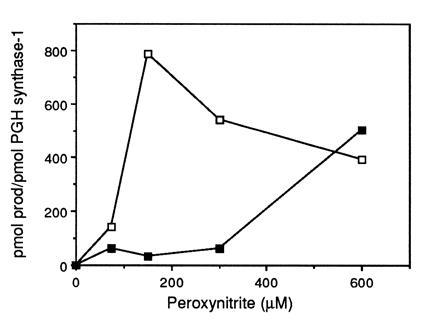
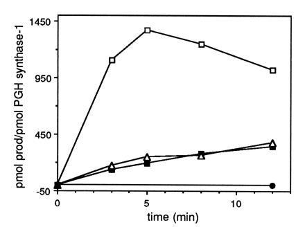
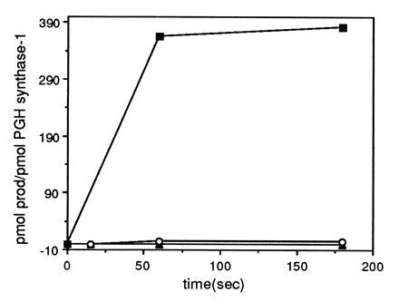
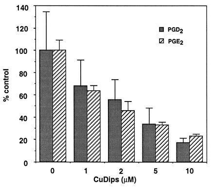
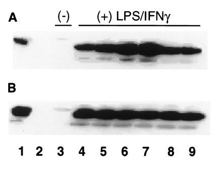
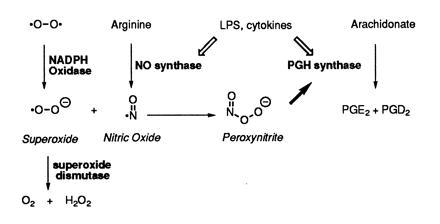
Peroxynitrite activates the cyclooxygenase activities of constitutive and inducible prostaglandin endoperoxide synthases by serving as a substrate for the enzymes' peroxidase activities. Activation of purified enzyme is induced by direct addition of peroxynitrite or by in situ generation of peroxynitrite from NO coupling to superoxide anion. Cu,Zn-superoxide dismutase completely inhibits cyclooxygenase activation in systems where peroxynitrite is generated in situ from superoxide. In the murine macrophage cell line RAW264.7, the lipophilic superoxide dismutase-mimetic agents, Cu(II) (3,5-diisopropylsalicylic acid)2, and Mn(III) tetrakis(1-methyl-4-pyridyl)porphyrin dose-dependently decrease the synthesis of prostaglandins without affecting the levels of NO synthase or prostaglandin endoperoxide synthase or by inhibiting the release of arachidonic acid. These findings support the hypothesis that peroxynitrite is an important modulator of cyclooxygenase activity in inflammatory cells and establish that superoxide anion serves as a biochemical link between NO and prostaglandin biosynthesis.


Figures

Figure 1
Figure 1
Peroxynitrite stimulation of AA metabolism by PGH synthase-1. Purified ram seminal vesicle PGH synthase-1 (22 nM) was incubated at 37°C with 8 units bovine erythrocyte GSH-Px, GSH, and 10 μg catalase in 0.1 M NaPO4 (pH 8.0) containing 500 μM phenol (200 μl reaction volume). Following addition of 50 μM [1-14C]AA, peroxynitrite was added and reactions were terminated with ethyl ether/methanol/1 M citric acid (pH 4.0) (30:4:1) after 2 min. Radiolabeled arachidonate metabolites were separated by thin-layer chromatography and quantitated using a radioactivity scanner. Control PGH synthase-1 activity in the absence of GSH-Px was 870 and 559 pmol product/pmol PGH synthase-1 at 0.2 and 1 mM GSH, respectively. □, 0.2 mM GSH; ▪, 1 mM GSH.
Figure 2
Figure 2
SIN-1 activation of GSH-Px/GSH inhibited PGH synthase-1. The assay conditions were identical to those described in the legend to Fig. 1 except that SIN-1 (750 μM) was added instead of peroxynitrite. Control PGH synthase-1 activity in the absence of GSH-Px was 1155 and 813 pmol product/pmol PGH synthase-1 at 0.25 and 2.5 mM GSH, respectively. □, 0.25 mM GSH; ▪, 1.0 mM GSH; ▵, 2.5 mM GSH; •, 0.25 mM GSH plus 1 μM SOD. The rate of formation of peroxynitrite from 750 μM SIN-1 in 0.1 M NaPO4 (pH 8.0) containing 500 μM phenol was estimated to be 30 μM/min following a 60-sec lag by monitoring the rate of oxygen uptake.
Figure 3
Figure 3
SNAP plus xanthine/xanthine oxidase activation of GSH-Px/GSH inhibited PGH synthase-1. PGH synthase-1 (75 nM) was incubated at 37°C in 1.3 ml 0.1 M NaPO4 (pH 8.0) containing 500 μM phenol, 0.25 mM GSH, and 120 units GSH-Px. Following addition of 50 μM [1-14C]AA, 300 μM SNAP, and 100 μM xanthine plus 0.2 unit xanthine oxidase or both were added. Aliquots of the reaction mixture were removed at the indicated time points and terminated with ethyl ether/methanol/1 M citric acid (pH 4.0) (30:4:1). Radiolabeled prostaglandin products were quantitated as described in Fig. 1. Control PGH synthase-1 activity in the absence of GSH-Px was 395 pmol product/pmol PGH synthase-1. ▴, 300 μM SNAP; ○, 100 μM xanthine plus 0.2 unit xanthine oxidase; •, SNAP plus xanthine/xanthine oxidase.
Figure 4
Figure 4
Inhibition of prostaglandin formation in LPS/IFN-γ-treated RAW264.7 cells by CuDips. RAW264.7 cells (3.5 × 10⁶ cells/T25 flask) were activated with 500 ng/ml LPS and 10 units/ml IFN-γ in serum-free SMEM for 7 hr. The cell monolayers were washed with PBS at t = 7 hr and then incubated in fresh PBS containing 0–10 μM CuDips for 1 hr. The dimethyl sulfoxide vehicle was kept constant at 1% in all flasks. At t = 8 hr of activation, the PBS was removed and analyzed for prostaglandin content by gas chromatography/negative ion chemical ionization mass spectrometry (54). The activated cells secreted 138 ng PGD2 and 1.4 ng PGE2 per 106 cells. These values were taken as 100% conversion, respectively. At all concentrations of CuDips, there was a statistically significant difference in eicosanoid production.
Figure 5
Figure 5
Effect of CuDips or MnTMPyP on PGH synthase-2 protein levels in activated RAW264.7 cells. RAW264.7 cells (4.4 × 10⁶ cells/T25 flask) were activated with 500 ng/ml LPS and 10 units/ml IFN-γ in serum-free SMEM for 7 hr. The activation medium was replaced with 1 ml PBS ± 10 μM CuDips or 20 μM MnTMPyP for 15, 30, 45, or 60 min at 37°C, such that all incubations ended after a total of 1 hr in PBS. The 60-min washout controls were washed twice with PBS, followed by a 15-min incubation in PBS without superoxide scavenger. Cells were scraped off the flasks into microfuge tubes, and the pellets were frozen at −80°C. The pellets were resuspended in 180 μl 100 mM Tris·HCl (pH 8) and 0.25 M sucrose for 5 min, and heated 10 min at 95°C with 100 μl SDS/DTT sample buffer. Samples (25 μl) were run on 10% resolving PAGE and transferred to polyvinylidene difluoride membranes (Millipore). Blots were incubated with PGH synthase-2 antibody 569 (1:3000) as the primary antibody, followed by the Amersham ECL Western detection system and a 7-sec exposure. Lanes: 1, 62 ng recombinant human recombinant PGH synthase-2; 2, blank; 3, nonactivated cells; 4, 8-hr activated cells; 5, activated cells treated 15 min with scavenger; 6, 30-min treatment; 7, 45-min treatment; 8, 60-min treatment; 9, 60-min treatment with washout. (A) CuDips (10 μM). (B) MnTMPyP (20 μM).
Figure 6
Figure 6
Relation of NO, O2, and prostaglandin biosynthesis in inflammatory cells.

References

    1. Needleman P, Turk J, Jakschik B A, Morrison A R, Lefkowith J B. Annu Rev Biochem. 1986;55:69–102. - PubMed
    1. Smith W L, Marnett L J. In: Metal Ions in Biological Systems. Sigel H, Sigel A, editors. Vol. 30. Basel: Dekker; 1994. pp. 163–199.
    1. Hamberg M, Svensson J, Wakabayashi T, Samuelsson B. Proc Natl Acad Sci USA. 1974;71:345–349. - PMC - PubMed
    1. Nugteren D H, Hazelhof E. Biochim Biophys Acta. 1973;326:448–461. - PubMed
    1. Vane J, Botting R. FASEB J. 1988;2:89–96. - PubMed

Publication types

MeSH terms

LinkOut - more resources