Comparative Study
. 1996 Dec 24;93(26):15108-13.
doi: 10.1073/pnas.93.26.15108.

Direct interaction of flagellin termini essential for polymorphic ability of flagellar filament

Affiliations
Comparative Study

Direct interaction of flagellin termini essential for polymorphic ability of flagellar filament

Y Mimori-Kiyosue et al. Proc Natl Acad Sci U S A. .

Abstract

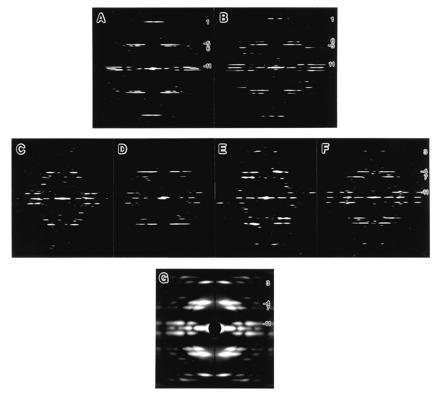
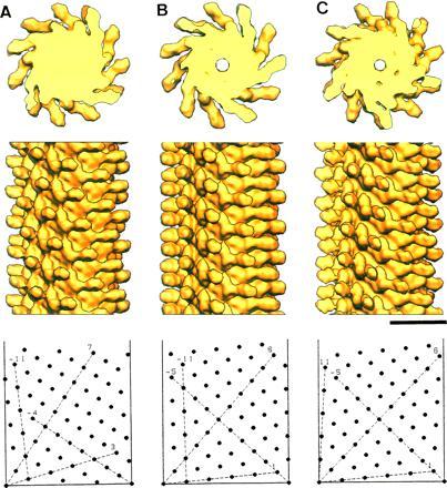
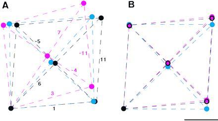
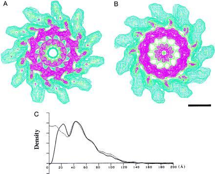
We report the structures of flagellar filaments reconstituted from various flagellins with small terminal truncations. Flagellins from Salmonella typhimurium strains SJW1103 (wild type), SJW1660, and SJW1655 were used, which form a left-handed supercoil, the L- and R-type straight forms, respectively. Structure analyses were done by electron cryomicroscopy and helical image reconstruction with a help of x-ray fiber diffraction for determining precise helical symmetries. Truncation of either terminal region, irrespective of the original flagellin species, results in a straight filament having a helical symmetry distinct either from the L- or R-type. This filament structure is named Lt-type. Although the local subunit packing is similar in all three types, a close comparison shows that the Lt-type packing is almost identical to the R-type but distinct from the L-type, which demonstrates the strong two-state preference of the subunit interactions. The structure clearly suggests that both termini are located in the inner tube of the concentric double-tubular structure of the filament core, and their proper interaction is responsible for the correct folding of fairly large terminal regions that form the inner tube. The double tubular structure appears to be essential for the polymorphic ability of flagellar filaments, which is required for the swimming-tumbling of bacterial taxis.


Figures

Figure 1
Figure 1
Computed Fourier transforms and x-ray diffraction pattern of various reconstituted filaments. (AF) Computed Fourier transforms of electron cryomicrographs of frozen hydrated filaments: (A) SJW1660 (the L-type), (B) SJW1655 (the R-type), (C) F-(1–486) of SJW1103 (wild type), (D) F-(1–486) of SJW1660, (E) F-(1–486) of SJW1655; and (F) F-(20–494) of SJW1655. (G) X-ray fiber diffraction pattern from an oriented filament sol of SJW1655 F-(1–486). For all the figures, the data out to about 20-Å resolution are shown. The order of Fourier–Bessel component (or helical start number), n, of each layer line is indicated on the right side. In the Fourier transforms of the L- and R-type (A and B), relative layer-line intensities and the order of Fourier–Bessel component are strongly correlated because the local subunit packing is similar to each other, and this makes it easy to identify the order. Therefore, the difference in helical symmetry between the two, such as the tilt of the 11-start helix, can be roughly identified from the axial positions of the major layer lines. If the strong layer line, n = −5, is above the relatively weaker layer line, n = 6, the 11-stranded protofilaments are tilted to the left, or vice versa. The larger the distance between the two layer lines, the larger the tilt angle (see also Fig. 2 B and C). This relation is also valid in the Fourier transforms of the filaments of truncated flagellins (CF), except that some of the Fourier–Bessel components have slightly different orders: e.g., n = 1 of the L- and R-type is 3; n = −5 is −4; n = 6 is 7; and so on. This indicates that the local packing and orientation of the subunits are more or less preserved also in the filaments of truncated flagellins.
Figure 2
Figure 2
Solid surface representation of the reconstructed density maps. F-(1–486) of SJW1103 (A), SJW1660 (B), and SJW1655 (C). (Top) Axial view of 50-Å-thick cross sections showing 11 subunits in two turns of one-start helix. (Middle) Side view of 300-Å-long segments. (Bottom) Helical lattice showing subunit packing arrangements at the radius of 45 Å, where the radial density distribution of the outer tube has its peak (see Fig. 3C). The data out to 12-Å resolution are included for calculating the density maps. The numbers of averaged filament images for reconstruction are 20, 14, and 16, and average phase residuals of individual images to the reference images are 17 (±4), 21 (±7), and 14 (±3) degree, for A, B, and C, respectively. (Bar = 100 Å.)
Figure 4
Figure 4
Comparison of the local subunit packing of the three different helical symmetries. (A) Local lattices of the Lt-, L-, and R-type are superimposed with the filament axis vertical. (B) Lattices are tilted so that the lattice lines representing the protofilaments (the 11-start) are superimposed. Lines and circles are colored red for the Lt-type, blue for the L-type, and black for the R-type. The lattices are drawn on the cylindrical surface at the radius of 45 Å, which corresponds to the radial position of the center of the outer-tube structure shown in Fig. 3. The lattice parameters were calculated from the helical symmetries (Table 1), which were obtained from the layer-line spacings in the x-ray fiber diffraction patterns (the diffraction pattern of the Lt-type is in Fig. 1G; data not shown for the L- and R-type). (Bar = 20 Å.)
Figure 3
Figure 3
Wire frame representation of the density map of the filaments showing higher contour levels and their radial density distributions. (A) SJW1655. (B) F-(1–486) of SJW1103. Both maps are axial views of 50-Å-thick cross sections. Color coding of contour lines indicates density levels of 1.0, blue; 1.8, green; 2.1, red. The contour lines in blue approximately cover the correct molecular volume. (Bar = 50 Å). In A, the contour lines in red show a concentric double-tubular structure in the core of the filament, which is a common feature of all the intact filaments of supercoiled, the L- and R-type straight forms. In B, the density corresponding to the inner tube moved toward the axis, while the other part remained unchanged. (C) Radial density distributions of the filaments of intact and truncated flagellins. The solid line represents the density distribution of SJW1655, and the dotted line represents that of F-(1–486) of SJW1103.

Similar articles

Cited by

References

    1. Macnab R M, Ornston M K. J Mol Biol. 1977;112:1–30. - PubMed
    1. Larsen S H, Reader R W, Kort E N, Tso W W, Adler J. Nature (London) 1974;249:74–77. - PubMed
    1. Berg H C, Anderson R A. Nature (London) 1973;245:380–382. - PubMed
    1. Silverman M, Simon M. Nature (London) 1974;249:73–74. - PubMed
    1. Asakura S. Adv Biophys. 1970;1:99–155. - PubMed

Publication types

LinkOut - more resources