. 1996 Dec 24;93(26):15146-51.
doi: 10.1073/pnas.93.26.15146.

Identification of fission yeast nuclear markers using random polypeptide fusions with green fluorescent protein

Affiliations

Identification of fission yeast nuclear markers using random polypeptide fusions with green fluorescent protein

K E Sawin et al. Proc Natl Acad Sci U S A. .

Abstract

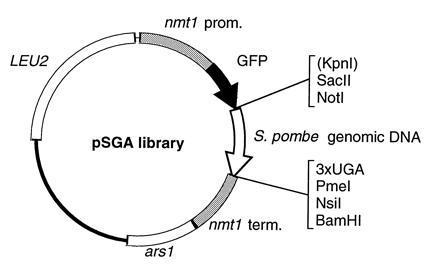
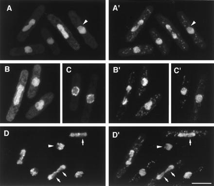
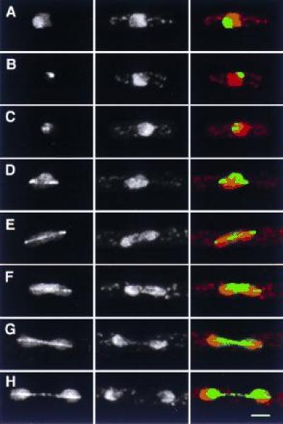
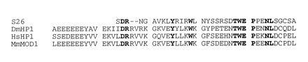
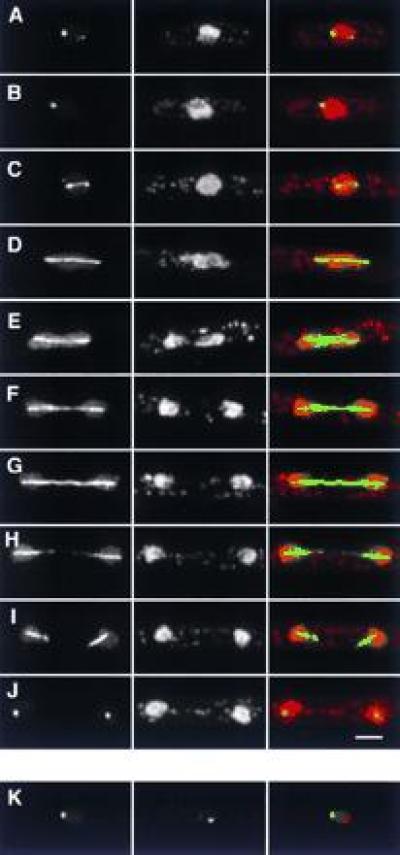
We describe a method for identifying genes encoding proteins with stereospecific intracellular localizations in the fission yeast Schizosaccharomyces pombe. Yeast are transformed with a gene library in which S. pombe genomic sequences are fused to the gene encoding the Aequorea victoria green fluorescent protein (GFP), and intracellular localizations are subsequently identified by rapid fluorescence screening in vivo. In a model application of these methods to the fission yeast nucleus, we have identified several novel genes whose products are found in specific nuclear regions, including chromatin, the nucleolus, and the mitotic spindle, and sequence similarities between some of these genes and previously identified genes encoding nuclear proteins have validated the approach. These methods will be useful in identifying additional components of the S. pombe nucleus, and further extensions of this approach should also be applicable to a more comprehensive identification of the elements of intracellular architecture in fission yeast.


Figures

Figure 1
Figure 1
Diagram of the pSGA plasmid library used for screening (see Materials and Methods). Shown flanking the genomic DNA insert are unique restriction sites (KpnI is non-unique) and three stop codons, each in a different reading frame.
Figure 2
Figure 2
GFP fusion proteins localized to the nucleus. (AD) GFP fluorescence; (A′–D′) corresponding images of DNA staining by PI. (A and A′) Localization of M31. (B and B′) Localization of M30 at low expression levels. (C and C′) Nuclear “rim” localization of M30 at higher expression levels (see text). (D and D′) Localization of M25 to nuclear chromatin. Note lack of staining in the interphase nucleolus, which stains weakly with PI (arrowhead), and lack of association of M25 with specific strands of segregating chromatin (arrows), which may be embedded in nucleolar material. (Bar = 5 μm.)
Figure 3
Figure 3
GFP fusion proteins with nucleolar localizations. (Left) GFP; (Center) DNA (PI stain); (Right) merged image of GFP (green) and DNA (red). (AE) General nucleolar localization of S32, during interphase (A) and different stages of mitosis (BE). (FH) Localization of S2 in interphase (F) and mitosis (G and H). Note the more compact organization of S2 in mitosis relative to S32. (IN) Punctate nucleolar localization of S19 in interphase (I and J) and mitosis (KM), and more general nucleolar localization when highly overexpressed (N). Note the specific association of S19 and trailing chromatin strands in L. (Bar = 2 μm.)
Figure 4
Figure 4
Localization of S26 to the nucleolus during interphase, and to the nucleolus and mitotic spindle during mitosis. (Left) GFP; (Center) DNA (PI stain); (Right) merged image of GFP (green) and DNA (red). (AC) Interphase cells. Note heterogeneity of fluorescence, which in all cases is confined to the nucleolus. (DH) Different stages of mitosis. During mitosis, S26 remains associated with nucleolar regions, albeit to varying degrees in different cells (compare E with F, and G with H). (Bar = 2 μm.)
Figure 5
Figure 5
Comparison of amino acid sequence from the beginning of clone S26 (from the fusion joint with GFP) with chromodomains from Drosophila melanogaster HP1 (GenBank accession no. M57574M57574), a human homolog of HP1 (accession no. L07515L07515), and mouse Modifier 1 protein (accession no. P23197P23197). Residues conserved among all four proteins are shown in bold. The alignment of the latter three sequences is based on that of Aasland and Stewart (24) and further details and comparisons with other chromodomain proteins can be found therein.
Figure 6
Figure 6
Localization of S5. (AJ) Localization in interphase and in different stages of mitosis. (Left) GFP; (Center) DNA (PI stain); (Right) merged image of GFP (green) and DNA (red). Note the fainter, secondary spots in both interphase (A) and early mitosis (C), the collapse of spindle localization into a single spot at the end of mitosis (J), and the lack of increased S5 fluorescence at spindle poles. (K) Comparison of S5 and spindle pole body localization. (Left) GFP; (Center) anti-sad1p; (Right) merged image of GFP (green) and anti-sad1p staining (red). The two signals are distinct. (Bar = 2 μm.)

References

    1. Hayles J, Nurse P. Annu Rev Genet. 1992;26:373–402. - PubMed
    1. Moreno S, Klar A, Nurse P. Methods Enzymol. 1991;194:795–823. - PubMed
    1. Nurse P. Nature (London) 1990;344:503–508. - PubMed
    1. Snell V, Nurse P. EMBO J. 1994;13:2066–2074. - PMC - PubMed
    1. Nurse P. Mol Biol Cell. 1994;5:613–616. - PMC - PubMed

Publication types

MeSH terms

LinkOut - more resources