. 1996 Dec 24;93(26):15158-63.
doi: 10.1073/pnas.93.26.15158.

The aphid transmission factor of cauliflower mosaic virus forms a stable complex with microtubules in both insect and plant cells

Affiliations

The aphid transmission factor of cauliflower mosaic virus forms a stable complex with microtubules in both insect and plant cells

S Blanc et al. Proc Natl Acad Sci U S A. .

Abstract

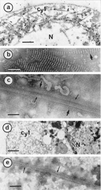
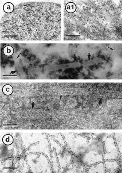
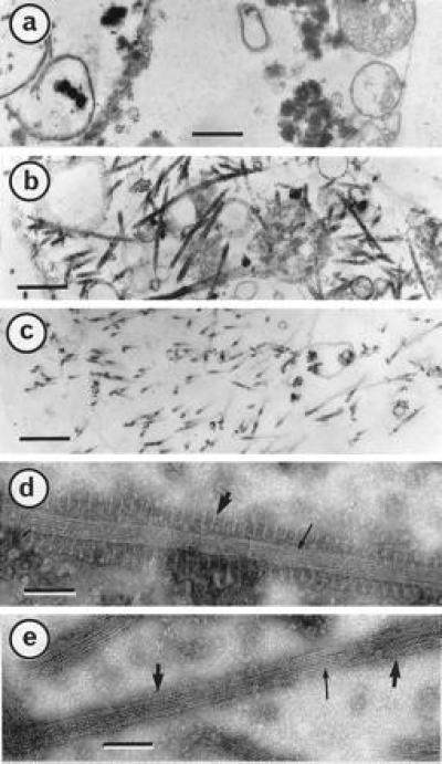
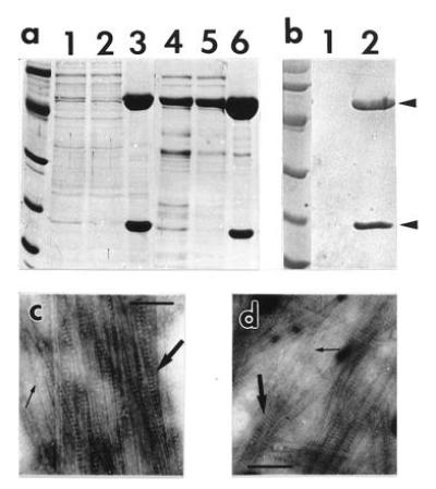
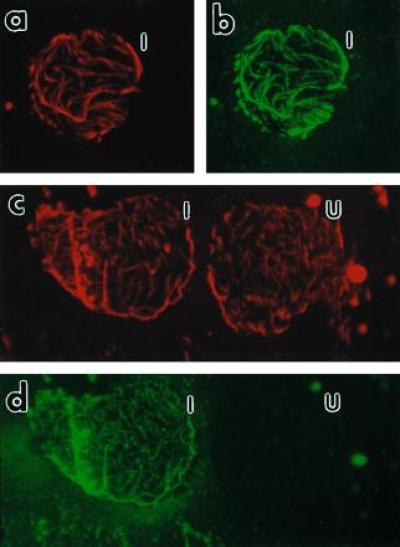
We analyzed the distribution of the cauliflower mosaic virus (CaMV) aphid transmission factor (ATF), produced via a baculovirus recombinant, within Sf9 insect cells. Immunogold labeling revealed that the ATF colocalizes with an atypical cytoskeletal network. Detailed observation by electron microscopy demonstrated that this network was composed of microtubules decorated with paracrystalline formations, characteristic of the CaMV ATF. A derivative mutant of the ATF, unable to self-assemble into paracrystals, was also analyzed. This mutant formed a net-like structure, with a mesh of four nanometers, tightly sheathing microtubules. Both the ATF- and the derivative mutant-microtubule complexes were highly stable. They resisted dilution-, cold-, and calcium-induced microtubule disassembly as well as a combination of all three for over 6 hr. CaMV ATF cosedimented with microtubules and, surprisingly, it bound to Taxol-stabilized microtubules at high ionic strength, thus suggesting an atypical interaction when compared with that usually described for microtubule-binding proteins. Using immunofluorescence double labeling we also demonstrated that the CaMV ATF colocalizes with the microtubule network when expressed in plant cells.


Figures

Figure 1
Figure 1
P18 paracrystals decorate MTs within Sf9 cells. Cells infected for 3 days were harvested and processed for electron microscopic observations and gold-labeling as described (15). (a) A thin section of an infected Sf9 cell was gold-labeled by a P18 antibody. As reference, negatively stained characteristic P18 paracrystals (15) are shown in b. (c) Infected cells were treated with Taxol and disrupted in AB buffer before negative staining. P18 paracrystals and well-shaped MTs are indicated with large and small arrows, respectively. (d) Thin section of a cell infected with a wild type baculovirus. (e) Negative staining of a similar cell disrupted after a Taxol treatment. Cyt, cytoplasm; N, nucleus. [Bars = 1 μm (a and d), 100 nm (b), and 75 nm (c and e).]
Figure 2
Figure 2
A P18 derivative mutant, P18 157m, is creating a net-like sheath on Sf9 cell MTs. Cells infected for 3 days with a baculovirus recombinant producing P18 157m were prepared for electron microscopic observations as in ref. 15. A thin section of an infected cell (a) was gold-labeled with a P18 antibody (a1). After being treated with Taxol, infected cells were disrupted in AB buffer, negatively stained, and observed (b and c). P18 157m net-like structure and well-shaped MTs are indicated by large and small arrows, respectively. (d) An infected cell was trapped on a microscope grid and gold labeled with a P18 antibody. (Bars = 500, 250, 75, 50, and 400 nm in a, a1, b, c, and d, respectively.)
Figure 3
Figure 3
P18– and P18 157m–MT complexes are highly stable. Cells were infected for 5 days, harvested, fixed and embedded as described (15). Micrographs represent thin sections of Sf9 cells infected with wild-type baculovirus (a), recombinant producing P18 (b), and recombinant producing P18 157m (c). P18– and P18 157m–MT complexes (d and e, respectively) were extracted and submitted to MT disassembly-inducing factors as described in the text before negative staining with ammonium molybdate (15). [Bars = 500 nm (ac) and 60 nm (d and e).]
Figure 4
Figure 4
Cosedimentation of P18 and MTs at elevated ionic strength. Samples were fractionated in a SDS/12% polyacrylamide gel and stained with Coomassie blue R250 (a) or Ponceau Red (Sigma) after transfer onto a nitrocellulose membrane (b). (a) Samples were processed as described in the text, and 10 μl were loaded in each lane. A salt-soluble P18 extract mixed with Sf9 preformed MTs was loaded in lane 1, and the supernatant and pellet (resuspended in 200 μl AB) from a 15,000 × g, 20-min centrifugation through a 15% sucrose cushion made in AB were loaded in lanes 2 and 3, respectively. P18 was also cosedimented with pig brain MTs. Samples in lanes 4, 5, and 6 were prepared in the same conditions as in lanes 1, 2, and 3, except for slight modifications indicated in the text. A control experiment (b) was carried out omitting the addition of preformed MTs in the salt-soluble P18 extract, and the corresponding pellet was loaded in lane 1; lane 2 is as in lane 3 in a. P18 and tubulin are indicated with arrows. Molecular weight reference scales are phosphorylase b (94 kDa), albumin (67 kDa), ovalbumin (43 kDa), carbonic anhydrase (30 kDa), trypsin inhibitor (20.1 kDa), and α-lactalbumin (14.4 kDa). Proteins of pellets loaded in lanes 3 and 6 in a were trapped on an electron microscope grid, negatively stained and observed (c and d, respectively). P18 paracrystals and MTs are indicated with large and small arrows. (Bars = 150 nm.)
Figure 5
Figure 5
Fluorescence micrographs of Nicotiana tabacum cv. Xanthi membrane ghosts. Membrane ghosts were double labeled as indicated for visualizing both MTs (a and c) and P18 (b and d). The same membrane ghost from an infected protoplast producing P18 is shown in a with a rhodamine filter cube for visualizing the MTs and in b with a fluorescein filter cube for visualizing P18. After Taxol treatment (see text), MTs of two membrane ghosts are shown as a rhodamine fluorescence in c and the presence of P18 in only one of these cells is shown by fluorescein in d. I, infected cell; U, uninfected cell. (×400 for ad.)

References

    1. Shepherd R J, Wakeman R J, Romanko R R. Virology. 1968;36:150–152. - PubMed
    1. Rothnie H M, Chapdelaine Y, Hohn T. Adv Virus Res. 1994;44:1–67. - PubMed
    1. Markham P G, Pinner M S, Raccah B, Hull R. Ann Appl Biol. 1987;111:571–587.
    1. Pirone T P. Semin Virol. 1991;2:81–87.
    1. Pirone T P, Blanc S. Annu Rev Phytopathol. 1996;34:227–247. - PubMed

MeSH terms

Substances

LinkOut - more resources