Review
. 1996 Dec 24;93(26):15195-202.
doi: 10.1073/pnas.93.26.15195.

On the molecular basis and regulation of cellular capacitative calcium entry: roles for Trp proteins

Affiliations
Review

On the molecular basis and regulation of cellular capacitative calcium entry: roles for Trp proteins

L Birnbaumer et al. Proc Natl Acad Sci U S A. .

Abstract

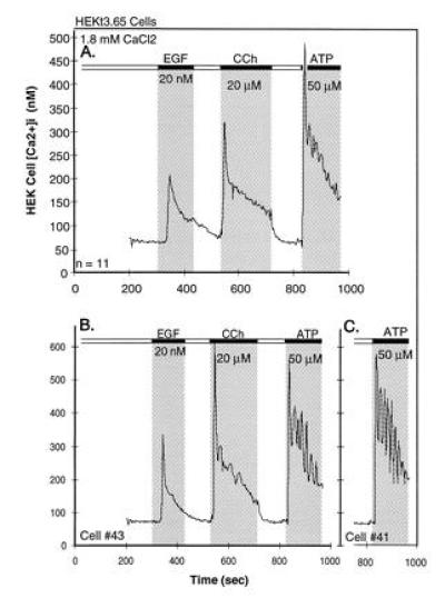
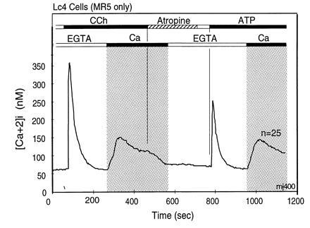
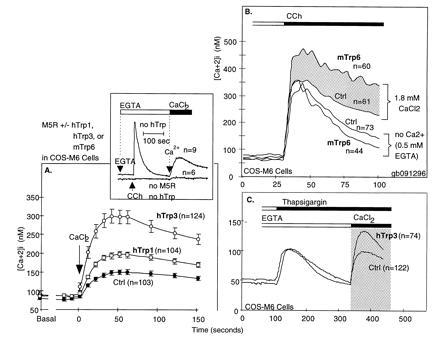
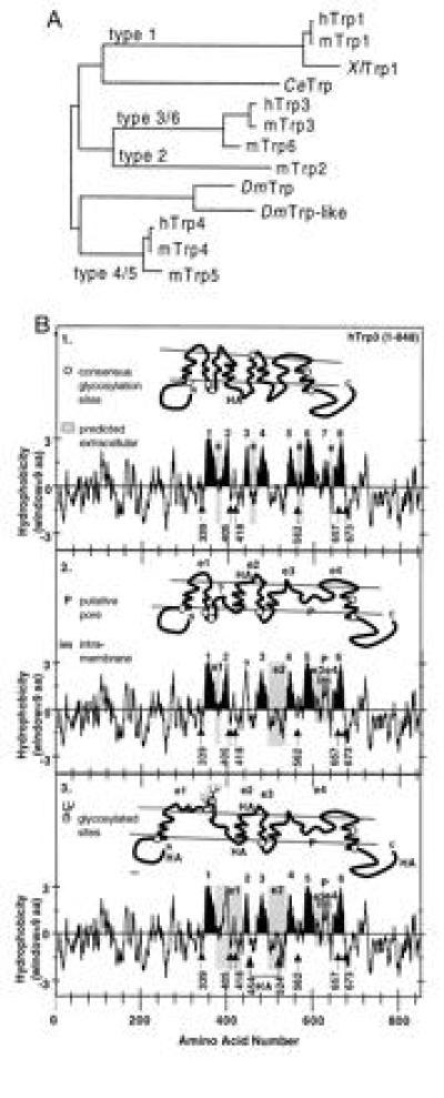
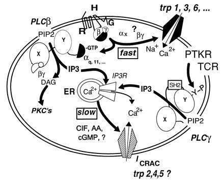
During the last 2 years, our laboratory has worked on the elucidation of the molecular basis of capacitative calcium entry (CCE) into cells. Specifically, we tested the hypothesis that CCE channels are formed of subunits encoded in genes related to the Drosophila trp gene. The first step in this pursuit was to search for mammalian trp genes. We found not one but six mammalian genes and cloned several of their cDNAs, some in their full length. As assayed in mammalian cells, overexpression of some mammalian Trps increases CCE, while expression of partial trp cDNAs in antisense orientation can interfere with endogenous CCE. These findings provided a firm connection between CCE and mammalian Trps. This article reviews the known forms of CCE and highlights unanswered questions in our understanding of intracellular Ca2+ homeostasis and the physiological roles of CCE.


Figures

Figure 1
Figure 1
[Ca2+]i transients in HEK-293 cells in response to epidermal growth factor, the muscarinic agonist CCh and the purinergic agonist ATP, as seen in the continuous presence of extracellular Ca2+. (A) Averaged changes in [Ca2+]i, observed in 11 cells. (B) Records of changes in two individual cells of the set. Additions and washouts of the agonists are depicted by the gray areas and the black bars at the top. For details see of [Ca2+]i determination see Zhu et al. (6).
Figure 2
Figure 2
Dissociation of agonist-stimulated [Ca2+]i transients into IP3-induced release and CCE-mediated replenishment components. The first rapid rise does not require extracellular Ca2+ and returns rapidly to the starting [Ca2+]i, due to extrusion of Ca2+ from the cells by plasma membrane Ca-ATPases and reuptake of Ca2+ into stores by sarcoplasmic endoplasmic reticulum Ca-ATPases. The second component, stimulation of CCE, is seen as influx of Ca2+ from the extracellular milieu that occurs upon Ca2+ addition after completion of the first Ca2+ transient. This form of Ca2+ entry is due to stimulation of CCE channels by a signal generated by the IP3-induced store depletion and/or by some other agonist-induced signaling process. [Ca2+]i changes were measured in murine L cells (Lc4 cells) expressing in stable form the M5 muscarinic acetylcholine receptor. Bars on top depict changes in composition of the medium. The lower bars indicate Ca2+ additions; open bar indicates addition of 0.5 mM EGTA to medium without added Ca2+, and the black bar and gray area depicts change to a balanced salt solution containing 1.8 mM CaCl2. The upper bars indicate agonist additions. When added, concentration of CCh was 20 μM and that of ATP was 90 μM. Note that the initial rise of [Ca2+]i after agonist addition in the absence of extracellular Ca2+ returns spontaneously to baseline levels, and also note that addition of extracellular Ca2+ uncovers the presence of an activated influx pathway that causes a robust increase in [Ca2+]i. This influx is referred to as CCE. Note further that in these cells the release of Ca2+ from intracellular stores occurs as a concerted event without hint of oscillations.
Figure 4
Figure 4
Functional expression of Trp proteins enhances CCE in COS Cells. (A) Enhancement of agonist-stimulated CCE by expression of hTrp3 in COS cells. Note that expression of the Trp protein caused an enhancement of CCE [adapted from Zhu et al. (6)]. (B) Rapid activation of mTrp6-induced Ca2+ influx occurs within the same time frame as IP3-induced Ca2+ release. Note that while, in the absence of external Ca2+, the initial IP3-mediated increase in [Ca2+]i in mTrp6-expressing cells is not significantly different from that seen in cells expressing the M5 receptor alone, the presence of external Ca2+ uncovers rapid stimulation of an additional flux of Ca2+ into the cytoplasm, which we interpret to come from the extracellular space and entering through CCE channels formed of Trp subunits. (C) Trp-based CCE channels are stimulated by store depletion. The figure shows agonist-independent activation of CCE by thapsigargin-induced depletion of Ca2+ stores in COS cells and its enhancement by either wild-type hTrp3.
Figure 3
Figure 3
Molecular diversity of 12 Trp sequences as deduced from their corresponding cDNAs and early speculations on secondary structure of CCE channels. (A) Phylogenetic tree obtained by pair-wise comparison of the amino acid sequences that span the putative pore region and the two putative transmembrane segments that flank this region (≈170 amino acids). This assignment assumes a transmembrane topology homologous to that of voltage-gated Ca2+, Na+, and K+ channels (B2 and 3). (B) Kyte–Doolittle plots and models of transmembrane topology of hTrp3 highlighting in black eight or two versions with six hydrophobic regions that may form transmembrane segments based on a possible homologous relation to voltage-gated ion channels. The location of consensus NXS/T glycosylation sites are highlighted (numbers correspond to the asparagines of the NXS/T motifs). In the six-transmembrane models, one of the hydrophobic regions thought not to span the membrane completely, P, is believed to be intramembrane (im) and to form the putative SS1-2 pore region used for the construction of the phylogenetic tree shown in A. e and e1-e4 are sequences predicted by the various models to be exposed to the outside of the cell. Note that, in B2, none of the six consensus glycosylation sites (○) is predicted to be glycosylated, while, in B3, two sites are exposed and presumed to be glycosylated. Locations at which HA epitopes were introduced are also shown.
Figure 5
Figure 5
Development of a nonselective cation current in HEK 293 cells expressing human Trp3 after loading with BAPTA. HEK cells were transfected with pcDNA3 carrying either the HA epitope-tagged Trp3 cDNA (hTrp3), or as control, the human type-2 vasopressin receptor cDNA (V2R) using the calcium phosphate/glycerol shock method. G418-resistant colonies were selected and tested for expression of the respective inserts—the HA epitope by immunocytochemistry and the V2R by its ability to stimulate adenylyl cyclase. Clonal cell lines were then isolated by limiting dilution, retested for expression of the proteins, and tested for ionic transmembrane currents using the whole-cell patch clamp technique. The pipette contained 150 mM Cs-aspartate, 4 mM MgCl2, 12 mM Na-BAPTA, and 10 mM Hepes. The extracellular medium contained normal saline: 155 mM NaCl, 2 mM KCl, 2 mM MgCl2, 10 mM glucose, and 10 mM Hepes. Na-free saline contained: 110 mM N-methylglucammonium (NMG)-Mes, 5 mM H-EDTA, 4.3 mM CaCl2, and 10 mM Hepes (free Ca2+, 20 μM). The cells were subjected to a depolarizing ramp from −150 mV to +150 mV. The upper portion shows the records of a control V2R-positive cell; the lower portion shows transmembrane currents of a cell expressing hTrp3. Note presence in the hTrp3-expressing cells of a nonselective conductance that is absent in control HEK cells as well as in the cell attached patch before establishing the whole cell configuration. Exchanging NMG+ for Na+ abolished the inward aspect of the transmembrane current but did not interfere with the outward current carried by Cs+ (data not shown). Other experiments showed that the channel is permeable to Ca2+ as well as Na+.
Figure 6
Figure 6
Expression of a 1.2-kb fragment of murine trp4 cDNA in antisense direction inhibits CCE stimulated by cotransfected M5 receptor. Murine L(tk) cells were transfected with cDNA encoding the M5 receptor alone (Fig. 2) or in combination with a 1.2-kb mTrp4 cDNA fragment transcribed in the antisense direction. Cells were selected for acquisition of G418 resistance encoded in the pcDNA3 vector that carried the M5 receptor cDNA. Lat4 cells were then analyzed for agonist-stimulated Ca2+ transients by video microscopy (6). The figure shows averaged records of changes in [Ca2+]i in Lat4 cells that responded to 20 μM CCh in the absence of external Ca2+ with a typical IP3-induced transient increase in [Ca2+]i, as seen in the absence of extracellular Ca2+. This was followed first by addition of 1.8 mM Ca2+in the presence of CCh, and then of 90 μM ATP in the presence of Ca2+. The addition of external Ca2+ tested for CCh-stimulated CCE and that of ATP for depletion of stores by CCh. Note that CCh indeed depleted the bulk of the intracellular stores, as seen by the failure of the saturating concentration of ATP to elicit a release of Ca2+. Note further that, in spite of full depletion of internal stores, CCE was not activated, indicating interference of the antisense cDNA with expression of one or more components required for CCE.
Figure 7
Figure 7
Glycosylation and immunocytochemical analysis of Trp3 suggests the transmembrane topology shown in Fig. 3B3. HEKt3-9 cells expressing HA epitope-tagged hTrp3 in stable form or COS-M6 cells transiently expressing the same Trp, were metabolically labeled with [35S]Met/Cys. Cells were solubilized, and the HA-tagged proteins were immunoprecipitated by incubation with C12A5 monoclonal antibody and protein A-Sepharose, eluted with HA peptide, and either treated or not treated with Endo H or PN-glycosidase F as described in ref. 44. The resulting samples were analyzed by 9% SDS/PAGE, followed by autoradiography. The figure shows a digitally acquired picture of the autoradiogram processed with the aid of photofinish and powerpoint softwares and printed with a Canon CJ10 printer. Note that untreated [35S]hTrp3 runs as a complex set of bands that are converted to a single band of ≈97 kDa by treatment with PN glycosidase F, indicating that protein was glycosylated by the HEK cells.
Figure 8
Figure 8
Models of secondary and quaternary structures of CCE channels and their subunits. CCE channels are hypothesized to be made of different Trps and/or different Trp combinations to account for CCE channels with varying ion selectivities and different forms of activation—e.g., store depletion-sensitive vs. store depletion-insensitive. The transmembrane topology chosen is arbitrarily based on that of voltage-gated K+, Na+, and Ca2+ channels and is thus far supported by the finding that hTrp3 is glycosylated and the results on HA localization in expression experiments. However, other transmembrane topologies are possible and the model may need major revisions. Existence of six nonallelic genes encoding six Trp-related proteins, opens the possibility that there are either diverse homomultimeric or diverse heteromultimeric CCE channels that could account for the functional heterogeneity of CCE seen in different tissues and cells.
Figure 9
Figure 9
Current hypotheses on the molecular makeup of CCE channels and their regulation. Multiple regulatory pathways can be identified. (i) The activated form of a G protein (Gq or other) directly activates a nonselective CCE channel encoded in Trp proteins, in analogy to activation of Drosophila Trp and Trpl by light. (ii) An activated α subunit of the Gq family or the βγ dimer of any G protein activated by receptor stimulation, stimulate PLCβ to form IP3 and diacylglycerol (DAG). DAG activates cellular PKCs and IP3 promotes Ca2+ release from internal stores through an IP3 receptor, the IP3-activated Ca2+ release channel of the endoplasmic reticulum. Depleted stores then signal to CCE channels of the nonselective and the Ca2+-selective type to allow entry of Ca2+. Whether highly Ca2+-selective CCE channels (ICRAC) are formed of Trps has not yet been determined. Likewise, whether ICRAC channels are regulated both by store depletion and an activated G protein(s) has not been studied. (iii) Receptors coupled by tyrosine kinases either through association with protein tyrosine kinases (PTKs) or by being themselves PTKs, as are the epidermal growth factor and insulin receptors, promote phosphorylation of PLCγ enzymes on tyrosines and thus cause their activation. IP3 formed in this way depletes Ca2+ stores and thus initiates activation of CCE channels. The nature of the store-to-CCE channel signal is not known.

References

    1. Birnbaumer L, Campbell K P, Catterall W A, Harpold M M, Hofmann F, Horne W A, Mori Y, Schwartz A, Snutch T P, Tanabe T, Tsien R W. Neuron. 1994;13:505–506. - PubMed
    1. Perez-Reyes E, Schneider T. Kidney Int. 1995;48:1111–1124. - PubMed
    1. Seeburg P H. Trends Neurosci. 1993;16:359–365. - PubMed
    1. Fasolato C, Innocenti B, Pozzan T. Trends Pharmacol Sci. 1994;15:77–83. - PubMed
    1. Striggow F, Ehrlich B E. Curr Opin Cell Biol. 1996;8:490–495. - PubMed

Publication types

Substances

LinkOut - more resources