. 1996 Dec 24;93(26):15209-14.
doi: 10.1073/pnas.93.26.15209.

DNA damage and p53-mediated cell cycle arrest: a reevaluation

Affiliations

DNA damage and p53-mediated cell cycle arrest: a reevaluation

N S Pellegata et al. Proc Natl Acad Sci U S A. .

Abstract

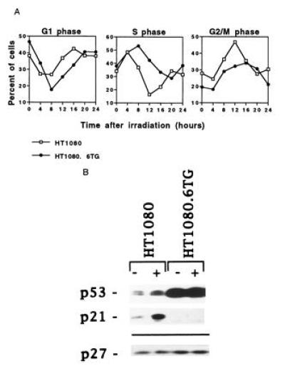
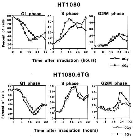
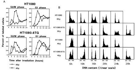
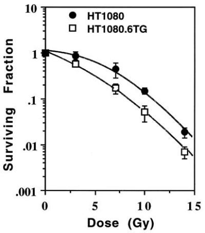
Most mammalian cells exhibit transient delays in the G1 and G2 phases of the cell cycle after treatment with radiation or radiomimetic compounds. p53 is required for the arrest in G1, which provides time for DNA repair. Recently, a role of p53 in the G2/M transition has also been suggested. However, it has been reported that the presence of functional p53 does not always correlate with the induction of these checkpoints. To precisely assess the role of p53 in activating cell cycle checkpoints and in cell survival after radiation, we studied the response of two isogenic human fibrosarcoma cell lines differing in their p53 status (wild type or mutant). We found that when irradiated cells undergo a wild-type p53-dependent G1 arrest, they do not subsequently arrest in G2. Moreover, wild-type p53 cells irradiated past the G1 checkpoint arrest in G2 but do not delay in the subsequent G1 phase. Furthermore, in these cell lines, which do not undergo radiation-induced apoptosis, the wild-type p53 cell line exhibited a greater radioresistance in terms of clonogenic survival. These results suggest that the two checkpoints may be interrelated, perhaps through a control system that determines, depending on the extent of the damage, whether the cell needs to arrest cell cycle progression at the subsequent checkpoint for further repair. p53 could be a crucial component of this control system.


Figures

Figure 1
Figure 1
Response to ionizing radiation of exponentially growing cells. (A) Flow cytometric analysis of HT1080 (□) and HT1080.6TG (•) cells treated with 4 Gy γ-radiation. The number of cells in S phase was determined by BrdUrd incorporation. The number of cells in G1 and G2/M phases was estimated from the flow histograms using PI fluorescence. The experiments were repeated at least twice per cell line and the averages are shown. (B) Western blot analysis of p53, p21, and p27 protein levels in control and irradiated cells from each tumor cell line. The levels of these proteins were detected by immunoblotting 4 hr after treatment with 4 Gy γ-rays (+) or following no treatment (−). The membrane was probed for p27 to check for equal loading since its expression does not change in response to radiation.
Figure 2
Figure 2
Cell cycle progression of synchronized cells irradiated in early G1 with 4 Gy of γ-radiation. (Upper) The percentage of control (0 Gy) or irradiated (4 Gy) synchronized HT1080 cells in the various phases of the cell cycle as a function of time after irradiation. (Lower) The percentage obtained for control (0 Gy) or irradiated (4 Gy) synchronized HT1080.6TG cells as a function of time.
Figure 3
Figure 3
Progression of BrdUrd-labeled cells through the cell cycle following treatment with 4 Gy of γ radiation of asynchronously growing (A) or synchronously growing (B) cells. (A) (Upper) The percentage of BrdUrd-labeled control (0 Gy) or irradiated (4 Gy) HT1080 asynchronous cells in G2/M and G1 phases as a function of time after irradiation. (Lower) The percentage of BrdUrd-labeled control (0 Gy) or irradiated (4 Gy) for asynchronous HT1080.6TG cells. The cells were pulsed with BrdUrd, and then the BrdUrd-positive population was followed throughout the cell cycle by FACS analysis gating to consider only the BrdUrd-positive cells. The values reported are the means of two independent experiments. (B) Flow cytometric profiles of BrdUrd-positive synchronized HT1080 or HT1080.6TG cells irradiated in early S (4 Gy) or left untreated (0 Gy). Cells were pulsed with BrdUrd when they were beginning to enter into the S phase and immediately irradiated. Cells were collected every 2 hr over a period of 32 hr and two-color FACS analysis performed gating for BrdUrd-positive cells.
Figure 4
Figure 4
Surviving fraction as a function of dose for HT1080 (•) and HT1080.6TG (□) cells γ-irradiated in exponential growth phase. Points, means of five flasks per dose in two independent experiments; bars = standard deviation.

Similar articles

Cited by

References

    1. Hartwell L H, Weinert T A. Science. 1989;246:629–634. - PubMed
    1. Hartwell L H, Kastan M B. Science. 1994;266:1821–1828. - PubMed
    1. Kastan M B, Onyekwere O, Sidransky D, Vogelstein B, Craig R. Cancer Res. 1991;51:6304–6311. - PubMed
    1. Kuerbitz S J, Plunkett B S, Walsh W V, Kastan M B. Proc Natl Acad Sci USA. 1992;89:7491–7495. - PMC - PubMed
    1. Clarke A R, Purdie C A, Harrison D J, Morris R G, Bird C C, Hooper M L, Wyllie A H. Nature (London) 1993;362:849–852. - PubMed

Publication types

Substances