. 1996 Dec 24;93(26):15215-20.
doi: 10.1073/pnas.93.26.15215.

Cyclin E, a redundant cyclin in breast cancer

Affiliations

Cyclin E, a redundant cyclin in breast cancer

J Gray-Bablin et al. Proc Natl Acad Sci U S A. .

Abstract

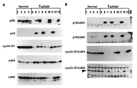
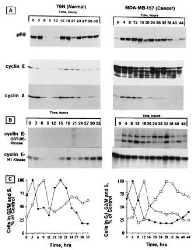
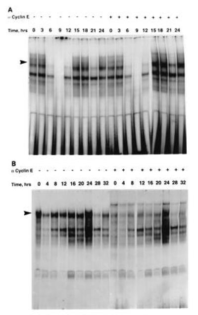
Cyclin E is an important regulator of cell cycle progression that together with cyclin-dependent kinase (cdk) 2 is crucial for the G1/S transition during the mammalian cell cycle. Previously, we showed that severe overexpression of cyclin E protein in tumor cells and tissues results in the appearance of lower molecular weight isoforms of cyclin E, which together with cdk2 can form a kinase complex active throughout the cell cycle. In this study, we report that one of the substrates of this constitutively active cyclin E/cdk2 complex is retinoblastoma susceptibility gene product (pRb) in populations of breast cancer cells and tissues that also overexpress p16. In these tumor cells and tissues, we show that the expression of p16 and pRb is not mutually exclusive. Overexpression of p16 in these cells results in sequestering of cdk4 and cdk6, rendering cyclin D1/cdk complexes inactive. However, pRb appears to be phosphorylated throughout the cell cycle following an initial lag, revealing a time course similar to phosphorylation of glutathione S-transferase retinoblastoma by cyclin E immunoprecipitates prepared from these synchronized cells. Hence, cyclin E kinase complexes can function redundantly and replace the loss of cyclin D-dependent kinase complexes that functionally inactivate pRb. In addition, the constitutively overexpressed cyclin E is also the predominant cyclin found in p107/E2F complexes throughout the tumor, but not the normal, cell cycle. These observations suggest that overexpression of cyclin E in tumor cells, which also overexpress p16, can bypass the cyclin D/cdk4-cdk6/p16/pRb feedback loop, providing yet another mechanism by which tumors can gain a growth advantage.


Figures

Figure 1
Figure 1
Expression and complex formation of p16/pRB pathway proteins in normal and tumor-derived breast epithelial cells. (A) Western blot analysis: exponentially growing normal and tumor cells were subjected to Western blot analysis using 50 μg of protein for each cell line in each lane of either a 6% (pRb), 13% (cyclin D1, cdk4, and cdk6), or 15% (p16) acrylamide gel and blotted as described. The same blot was reacted with cyclin D1, cdk4, and cdk6 affinity-purified antibodies. The blots were stripped between the three antibodies in 100 mM 2-mercaptoethanol, 62.5 mM Tris·HCl (pH 6.8), and 2% SDS for 30 min at 55°C. (B) Immune-complex formation: for immunoprecipitation followed by Western blot analysis, equal amounts of protein (500 μg) from cell lysates prepared from each cell line were immunoprecipitated with either monoclonal antibody to p16 (p16/cdk4 and p16/cdk6), polyclonal antibody to cyclin D1 (cyclin D1/cdk4), or a monoclonal antibody to cyclin D1 (cyclin D1/cdk6), coupled to protein A/G beads, and the immunoprecipitates were washed, boiled for 3 min, separated by SDS/13% PAGE, blotted to Immobilon membranes, and hybridized with either polyclonal antibody to cdk4 (p16/cdk4), polyclonal antibody to cdk6 (p16/cdk6 and cyclin D1/cdk6; arrow pointing to the complexed protein), or monoclonal antibody to cdk4 (cyclin D1/cdk4). The list of normal and tumor cell lines is presented in Table 1 using identical numbers.
Figure 2
Figure 2
Phosphorylation of pRb in synchronized population of tumor versus normal cells. Both cell types were synchronized by double thymidine block procedure. At the indicated times after release from double thymidine block, cell lysates were prepared and subjected to Western blot analysis (A) and histone H1 or GST-Rb kinase analysis (B). Protein (50 μg) for each time point was applied to each lane of either a 6% (pRb) or 10% (cyclins E and A) acrylamide gel and blotted as described. The same blot was reacted with cyclin E monoclonal (HE12) and cyclin A affinity-purified polyclonal antibodies. The blots were stripped between the two assays as described for Fig. 1. For kinase activity, equal amounts of proteins (600 μg) from cell lysates prepared from each cell line at the indicated times were immunoprecipitated with anti-cyclin E (polyclonal) coupled to protein A beads using either histone H1 or purified GST-Rb as substrates. (C) The relative percentage of cells in different phases of the cell cycle for each cell line at various times after release from double thymidine block was calculated from flow cytometric measurements of DNA content. ♦, cells in S phase; ○, cells in G2/M phase; □, cells in G1 phase.
Figure 3
Figure 3
Cyclin E is the predominant cyclin in p107/E2F complexes in tumor cells. E2F complexes were analyzed by gel retardation assays using cell lysates (15 μg) prepared from synchronized populations (see Fig. 2) of normal 76N cells (A) and tumor MDA-MB-157 cells (B). The oligonucleotide used as a labeled DNA probe includes the E2F binding site of the human dehydrofolate reductase promoter. The anti-cyclin E antibody (200 ng) was used to disrupt the E2F complexes.

References

    1. Hartwell L H, Kastan M B. Science. 1994;266:1821–1828. - PubMed
    1. Elledge S J, Harper J W. Curr Opin Cell Biol. 1994;6:847–852. - PubMed
    1. Morgan D O. Nature (London) 1995;374:131–134. - PubMed
    1. Sherr C J. Cell. 1994;79:551–555. - PubMed
    1. Nasmyth K. Curr Opin Cell Biol. 1993;5:166–179. - PubMed

Publication types

MeSH terms