. 1996 Dec 24;93(26):15299-304.
doi: 10.1073/pnas.93.26.15299.

A novel member of the RING finger family, KRIP-1, associates with the KRAB-A transcriptional repressor domain of zinc finger proteins

Affiliations

A novel member of the RING finger family, KRIP-1, associates with the KRAB-A transcriptional repressor domain of zinc finger proteins

S S Kim et al. Proc Natl Acad Sci U S A. .

Abstract

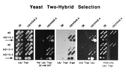
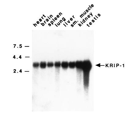
The Krüppel-associated box A (KRAB-A) domain is an evolutionarily conserved transcriptional repressor domain present in approximately one-third of zinc finger proteins of the Cys2-His2 type. Using the yeast two-hybrid system, we report the isolation of a cDNA encoding a novel murine protein, KRAB-A interacting protein 1 (KRIP-1) that physically interacts with the KRAB-A region. KRIP-1 is a member of the RBCC subfamily of the RING finger, or Cys3HisCys4, family of zinc binding proteins whose other members are known to play important roles in differentiation, oncogenesis, and signal transduction. The KRIP-1 protein has high homology to TIF1, a putative modulator of ligand-dependent activation function of nuclear receptors. A 3.5-kb mRNA for KRIP-1 is ubiquitously expressed among all adult mouse tissues studied. When a GAL4-KRIP-1 fusion protein is expressed in COS cells with a chloramphenicol acetyltransferase reporter construct with five GAL4 binding sites, there is dose-dependent repression of transcription. Thus, KRIP-1 interacts with the KRAB-A region of C2H2 zinc finger proteins and may mediate or modulate KRAB-A transcriptional repressor activity.


Figures

Figure 1
Figure 1
Selection of a KRAB-A interacting protein using a yeast two-hybrid screen. The second through the fifth panels demonstrate phenotyping on four media. Growth or LacZ expression reflects the expression levels of three GAL4-responsive reporter genes. Yeast transformants were patched in duplicate on SC-Leu-Trp (LeuTryp) medium in the panel on the left. The yeast cells were then replica plated onto the other four plates. In each panel are yeast cells transformed with pPC97/GAL4 encoding only the DNA binding (DB) region of GAL4 or pPC97/KRAB-A, which includes DB and KRAB-A (DB/KRAB-A), with the pPC86 vectors encoding one of the following: the activation domain (AD), AD and KRIP-1.3 (AD/15-3), or AD and KRIP-1.2 (AD/15.2). The arrows designate (from left to right on each panel) one negative control (DB + AD), one intermediate positive control (DB-retinoblastoma + AD-E2F1), and two positive controls (DB-Fos + AD-Jun, and full-length GAL4-containing binding domain and activation domain) (9). Protein–protein interaction confers ability to grow on HisLeuTryp medium in the presence of 50 mM 3-AT (second panel) and growth in UraTrypLeu medium (fourth panel). In the third, panel β-galactosidase activity induced by protein–protein interaction, is demonstrated. The fifth panel reveals FOA sensitivity resulting from the protein–protein interaction.
Figure 2
Figure 2
Nucleotide and protein sequence of KRIP-1. The RING finger, B1, B2, coiled coil domain, and PHD finger are boxed. In the case of the RING finger, B1, B2, and PHD finger region, the cysteines and histidines making up the consensus sequence are circled. In the case of the coiled coil domain, the amino acids that form the heptad repeat of hydrophobic amino acids are circled.
Figure 2
Figure 2
Nucleotide and protein sequence of KRIP-1. The RING finger, B1, B2, coiled coil domain, and PHD finger are boxed. In the case of the RING finger, B1, B2, and PHD finger region, the cysteines and histidines making up the consensus sequence are circled. In the case of the coiled coil domain, the amino acids that form the heptad repeat of hydrophobic amino acids are circled.
Figure 3
Figure 3
Multiple-organ Northern blot to detect KRIP-1 mRNA. Two micrograms of poly(A)-selected RNA from various organs was loaded onto each lane. The blot was hybridized at 65°C with the KRIP-1.2 cDNA and washed at 65°C in 0.1 × sodium chloride/sodium citrate (SSC)/0.1% SDS.
Figure 4
Figure 4
Coimmunoprecipitation of the KRAB-A domain from two distinct zinc finger proteins and KRIP-1.2. (A) COS cells were cotransfected with pMT3-KRIP-1.2, encoding the HA-tagged KRIP-1.2 with pMFH-Kid-1N, wild-type pMFH2, or pMFH2-Kid-1A, which encode T7–GAL4 fusion proteins with the non-zinc-finger region of Kid-1, no additional sequence, or the KRAB-A region of Kid-1 respectively. After cell lysis, preparation of soluble (S) and pellet (P) fractions, immunoprecipitation with the anti-HA antibodies, SDS/PAGE resolution of the S, P, and immunoprecipitate fractions (IP), transfer to an Immobilon-P membrane, and reaction with anti-T7 flag antibody, it is apparent that Kid-1N and Kid-1A immunoprecipitate with KRIP-1.2. (B) pMT3-KRIP-1.2 was cotransfected with pBXG1 constructs encoding GAL4 fusion proteins of Kid-1N, Kid-1A, and the KRAB-A region of ZNF2. Kid-1N and the KRAB-A regions of Kid-1 and ZNF2 coimmunoprecipitate with KRIP-1.2 when the complexes are precipitated by anti-HA antibody. The blot is stained with anti-GAL4 antibody.
Figure 5
Figure 5
Thin layer chromatography assays of CAT activities in COS cells cotransfected with a CAT reporter and with various KRIP-1 constructs. COS cells were cotransfected with 3 μg of pG5SV-BCAT reporter construct, which contains five GAL4 binding sites, and with 20 μg of the pBXG1 plasmid with inserts encoding a fusion protein of the DNA binding domain of GAL4 with Kid-1N antisense, full-length KRIP-1.2 encoding amino acids 43-834 of Krip-1, KRIP-1.2 in which the RING fingers, B1, B2, and coiled coil domains are deleted (GAL4-KRIP-1ΔRBCC), or the latter with a frameshift mutation resulting in a stop codon at nt 1727. Full-length KRIP-1.2 and KRIP-1.2 without the RBCC domains exert equivalent transcriptional repression. Numbers at the bottom reflect relative CAT activity.
Figure 6
Figure 6
Thin layer chromatography assays of CAT activities in COS cells cotransfected with a CAT reporter and with various amounts of full-length KRIP-1 constructs. The numbers at the top reflect the amount (in μg) of pMFH2/GAL4 plasmid encoding GAL4 binding domain only, pMFH2/GAL4-KRIP-1 encoding a GAL4-KRIP-1 fusion protein, and pMT3-KRIP-1 encoding an HA–KRIP-1 fusion protein that cannot bind to the GAL4 binding sites of pG5SV-BCAT. Numbers at the bottom reflect relative CAT activity.

Similar articles

Cited by

References

    1. Klug A, Rhodes D. Trends Biochem Sci. 1987;12:464–469.
    1. Bellefroid E J, Poncelet D A, Lecocq P J, Revelant O, Martial J A. Proc Natl Acad Sci USA. 1991;88:3608–3612. - PMC - PubMed
    1. Witzgall R, O’Leary E, Gessner R, Ouellette A J, Bonventre J V. Mol Cell Biol. 1993;13:1933–1942. - PMC - PubMed
    1. Witzgall R, O’Leary E, Leaf A, Önaldi D, Bonventre J V. Proc Natl Acad Sci USA. 1994;91:4514–4518. - PMC - PubMed
    1. Margolin J F, Friedman J R, Meyer W K-H, Vissing H, Thiesen H-J, Rauscher F J., III Proc Natl Acad Sci. 1994;91:4509–4513. - PMC - PubMed

Publication types

MeSH terms

Associated data