Comparative Study
. 1996 Dec 24;93(26):15335-40.
doi: 10.1073/pnas.93.26.15335.

Identification of a novel p53 functional domain that is necessary for efficient growth suppression

Affiliations
Comparative Study

Identification of a novel p53 functional domain that is necessary for efficient growth suppression

K K Walker et al. Proc Natl Acad Sci U S A. .

Abstract

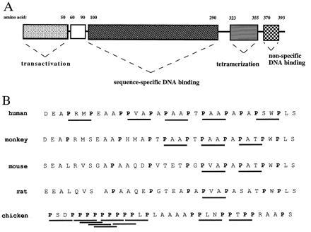
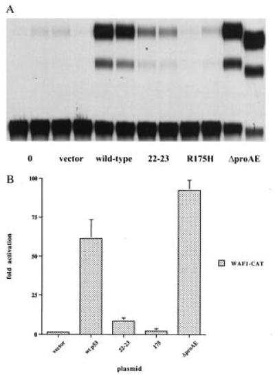
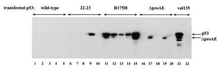
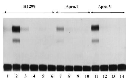
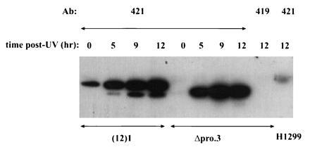
Activation of the p53 tumor suppressor protein has been demonstrated to block cell growth by inducing either a transient cell cycle arrest or programmed cell death (apoptosis). Although evidence exists linking p53's function as an activator of transcription to its ability to effect cell cycle arrest, the role of this activity in the induction of apoptosis remains unclear. To gain insight into the molecular mechanisms underlying p53-mediated antiproliferative pathways, a study was initiated to explore the functions of a putative p53 signaling domain. This region of the human p53 protein is localized between amino acids 61 and 94 (out of 393) and is noteworthy in that it contains five repeats of the sequence PXXP (where P represents proline and X any amino acid). This motif has been shown to play a role in signal transduction via its SH3 domain binding activity. A p53 cDNA deletion mutant (delta pro AE), which lacks this entire proline-rich domain (deleted for amino acids 62-91), was created and characterized for a variety of p53 functions. The entire domain has been shown to be completely dispensable for transcriptional activation. On the other hand, this deletion of the p53 proline-rich domain impairs p53's ability to suppress tumor cell growth in culture. Amino acid substitution mutations at residues 22 and 23 of p53 (eliminates transcriptional activity) also impair p53-mediated inhibition of cell growth in culture. Unlike wild-type p53, the delta proAE mutant cDNA can be stably expressed in tumor derived cell lines with few immediate detrimental effects. These cells express physiologic levels of p53 protein that are induced normally in response to DNA damage, indicating that removal of the proline-rich domain does not disrupt p53's upstream regulation by DNA damage. These data indicate that, in addition to the transcriptional activation domain, the p53 proline-rich domain plays a critical role in the transmission of antiproliferative signals down-stream of the p53 protein and may link p53 to a direct signal transduction pathway.


Figures

Figure 1
Figure 1
Structural features of the p53 protein. (A) Schematic representation of the domain structure of the human p53 protein indicating regions mediating transcriptional activation (amino acids 1–50), sequence-specific DNA binding (amino acids 100–290), tetramerization (amino acids 311–393), and non-specific DNA binding (amino acids 370–393). (B) Comparison of the predicted p53 amino acid sequence of the proline-rich domains from human (residues 61–94), monkey (residues 61–94), mouse (residues 55–88), rat (residues 60–92), and chicken (residues 56–89) as determined by Soussi et al. (37). Proline residues are indicated by boldface type and PXXP motifs are underlined.
Figure 2
Figure 2
Transcriptional transactivation activity of p53 mutants in H1299 cells. (A) Transactivation of the p53-responsive reporter, WAF1-CAT (21). cDNAs encoding for wild-type p53, the double point mutant 22-23, the tumor-derived mutant R175H, and the deletion mutant ΔproAE (deleted of residues 62–91) cloned into the mammalian expression vector, pRC-CMV (Invitrogen) were used for transactivation assays. Cells were cotransfected with 100 ng of p53 expression plasmid or the empty vector and 1 μg of reporter plasmid. Each transfection is presented in duplicate. (B) Quantitation of transactivation data displayed graphically as the fold activation of the WAF1-CAT reporter plasmid over its basal level of expression.
Figure 3
Figure 3
Analysis of H1299 cell lines stably transfected with mutant p53 plasmids. Clonal cell lines were generated by stable transfection with the plasmid pRC-CMV containing p53 cDNAs encoding the wild-type p53 protein (lanes 1–5), the transactivation mutant, 22-23 (lanes 6–10), the DNA binding mutant, R175H (lanes 11–15), the proline deletion mutant, ΔproAE (lanes 16–20), or the temperature-sensitive murine DNA binding mutant, A135V (lanes 21 and 22). Cell lines were analyzed for p53 protein expression by Western blot analysis of p53-specific immunocomplexes. Cell lysates were immunoprecipitated with the panspecific antibody, pAb421, which recognizes both mutant and wild-type conformations of p53 (lanes 1–21) or the negative control antibody, pAb419 (lane 22). Immunocomplexes were resolved by 10% SDS/PAGE, transferred to an Immobilon-P membrane (Millipore), and immunoblotted with pAb421. The positions of full-length p53 and the ΔproAE proteins are indicated.
Figure 4
Figure 4
Analysis of the ability of stably expressed ΔproAE to activate transcription. Transactivation of transiently transfected CAT reporter constructs. The H1299 cell line (lanes 1–6) and two independent derivatives stably expressing ΔproAE protein, Δpro.1 (lanes 7–10), and Δpro.3 (lanes 11–14), were transfected with 1 μg of the p53-responsive reporter WAF1-CAT (lanes 1–3, 7 and 8, and 11 and 12) or the negative control reporter Gal4-CAT (lanes 4–6, 9 and 10, and 13 and 14) in the presence (lanes 3, 6, 8, 10, 12, and 14) or absence (lanes 1, 2, 4, 5, 7, 9, 11, and 13) of 10 μg of the human MDM2 expression vector, HDM2. As a positive control for transactivation, 200 ng of the ΔproAE expression plasmid was transfected into the H1299 parental line (lanes 2 and 3).
Figure 5
Figure 5
UV induction of ΔproAE protein. The cell lines (12)1 (immortalized murine fibroblast expressing endogenous wild-type p53) and Δpro.3 (derivative of the tumor line H1299 stably expressing ΔproAE) were irradiated with a high dose of UV light (20 J/m2). Cells were collected at 0, 5, 9, and 12 hr posttreatment. Steady-state levels of p53 protein were determined by immunoprecipitation/Western analysis. Cell lysates were immunoprecipitated with the p53-specific monoclonal antibody, pAb421. Negative controls included precipitating the Δpro.3 12-hr lysate with the antibody pAb419, which does not recognize p53 and the p53-null H1299 lysate with pAb421. Immunocomplexes were resolved by 10% SDS/PAGE and transferred to an Immobilon membrane (Millipore). The membrane was probed with the p53-specific antibody pAb421 and bound antibody was detected with 125I-conjugated protein A. Data were quantitated using PhosphorImager analysis (Molecular Dynamics) and imagequant software. The positions of wild-type and ΔproAE p53 are indicated.

Similar articles

Cited by

References

    1. Hollstein M, Sidransky D, Vogelstein B, Harris C C. Science. 1991;253:49–53. - PubMed
    1. Maltzman W, Czyzyk L. Mol Cell Biol. 1984;4:1689–1694. - PMC - PubMed
    1. Kastan M B, Onyekwere O, Sidransky D, Vogelstein B, Craig R W. Cancer Res. 1991;51:6304–6311. - PubMed
    1. Lu X, Lane D P. Cell. 1993;75:765–778. - PubMed
    1. Lowe S W, Ruley H E. Genes Dev. 1993;7:535–545. - PubMed

Publication types

MeSH terms

LinkOut - more resources