. 1996 Dec 24;93(26):15370-5.
doi: 10.1073/pnas.93.26.15370.

Cell surface expression of the epithelial Na channel and a mutant causing Liddle syndrome: a quantitative approach

Affiliations

Cell surface expression of the epithelial Na channel and a mutant causing Liddle syndrome: a quantitative approach

D Firsov et al. Proc Natl Acad Sci U S A. .

Abstract

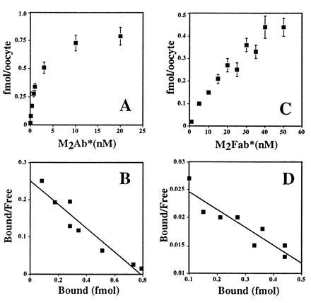
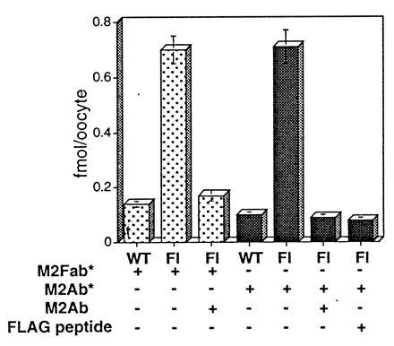
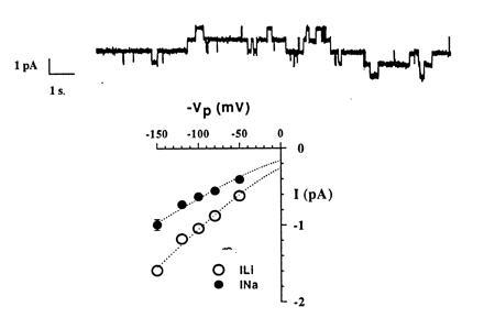
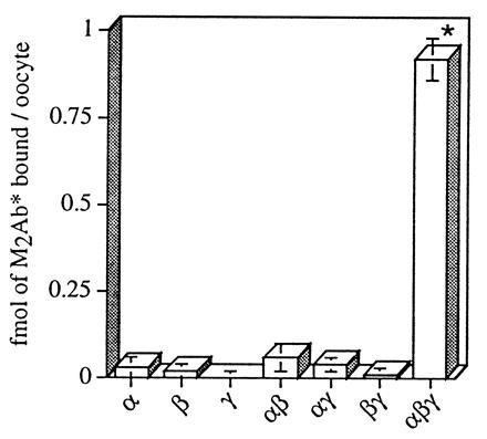
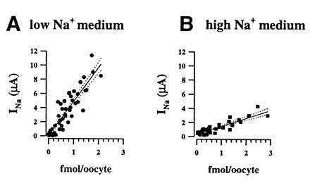
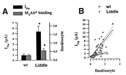
The epithelial amiloride-sensitive sodium channel (ENaC) controls transepithelial Na+ movement in Na(+)-transporting epithelia and is associated with Liddle syndrome, an autosomal dominant form of salt-sensitive hypertension. Detailed analysis of ENaC channel properties and the functional consequences of mutations causing Liddle syndrome has been, so far, limited by lack of a method allowing specific and quantitative detection of cell-surface-expressed ENaC. We have developed a quantitative assay based on the binding of 125I-labeled M2 anti-FLAG monoclonal antibody (M2Ab*) directed against a FLAG reporter epitope introduced in the extracellular loop of each of the alpha, beta, and gamma ENaC subunits. Insertion of the FLAG epitope into ENaC sequences did not change its functional and pharmacological properties. The binding specificity and affinity (Kd = 3 nM) allowed us to correlate in individual Xenopus oocytes the macroscopic amiloride-sensitive sodium current (INa) with the number of ENaC wild-type and mutant subunits expressed at the cell surface. These experiments demonstrate that: (i) only heteromultimeric channels made of alpha, beta, and gamma ENaC subunits are maximally and efficiently expressed at the cell surface; (ii) the overall ENaC open probability is one order of magnitude lower than previously observed in single-channel recordings; (iii) the mutation causing Liddle syndrome (beta R564stop) enhances channel activity by two mechanisms, i.e., by increasing ENaC cell surface expression and by changing channel open probability. This quantitative approach provides new insights on the molecular mechanisms underlying one form of salt-sensitive hypertension.


Figures

Figure 1
Figure 1
(A) FLAG-epitope placement into the rat ENaC sequence. The alignment of the extracellular loop sequences for the rat, human, and Xenopus ENaC subunits demonstrated the most important divergence in the region near the first transmembrane domain M1. This region was chosen to insert (α and γ subunits) or to replace (β subunit) the FLAG sequence (DYKDDDDK) at the indicated positions. (B) Immunoprecipitation of the FLAG-tagged ENaC subunits, using M2Ab*. Oocytes were injected with an α, β, or γ nontagged subunit alone (lane 1), or with an α, β, or γ FLAG-tagged subunit alone (lanes 2 and 3). Oocytes were metabolically labeled with [35S]methionine for a 4-h pulse followed by a 4-h chase period. Membranes were prepared, and proteins were immunoprecipitated and analyzed on 5–8% gradient SDS/PAGE. Immunoprecipitations of the samples corresponding to lane 3 were performed in the presence of 1000-fold molar FLAG peptide excess over the antibody concentration.
Figure 2
Figure 2
Equilibrium binding of M2Ab* and M2Fab* to the FLAG-tagged ENaC subunits. Stage V oocytes were injected, either with 10 ng of FLAG-tagged or with nontagged ENaC cRNAs. Twenty-four hours later, these oocytes were tested for M2Ab* (A) or M2Fab (C) binding. Twelve oocytes were transferred into 100 μl (M2Ab) or 50 μl (M2Fab) binding buffer and then incubated with increasing concentrations of M2Ab* or M2Fab* for 4 h on ice. The concentration of M2Ab* and M2Fab varied from 0.03–20 nM and 5–50 nM, respectively. Specific binding was calculated as the difference of the binding between the oocytes injected with FLAG-tagged and the oocytes injected with nontagged ENaC cRNAs and transformed into Scatchard plots for M2Ab* (B) or M2Fab* (D). (A and C) The means ± SEM of three independent experiments, each performed on 12 oocytes.
Figure 3
Figure 3
Specific binding of the M2Fab* and M2Ab* to Xenopus oocytes expressing FLAG-tagged ENaC subunits. Binding of the 50 nM M2Fab* (dotted columns) or 12 nM M2Ab* (shaded columns) was measured in parallel on the oocytes expressing either FLAG-tagged (Fl) or nontagged ENaC subunits. Specificity of the binding was tested, using either 1000-molar excess of the FLAG peptide (M2Fab* and M2Ab*) or 200-fold excess of unlabeled M2Ab (M2Ab*). FLAG peptide and unlabeled M2Ab were added in the binding solution simultaneously with the 125I-labeled agents. Shown are means ± SEM from four independent experiments, each performed on 12 oocytes.
Figure 4
Figure 4
Single-channel properties of FLAG-tagged ENaC channels. (Upper) A single-channel recording performed in cell attached configuration with Na+ ions as major cation in the pipette. Pipette voltage (Vpip) was 80 mV. Downward deflections represent channel openings. The four distinct current levels indicate the presence of at least four conducting channels in the patch. (Lower) Current-voltage relationship in the presence of Na+ or Li+ ions in the pipette. Single-channel conductance estimated from regression analysis between −50 mV and −150 mV was 5.5 pS for Na+ and 8.8 pS for Li+.
Figure 5
Figure 5
Specific M2Ab* binding to the oocytes injected with different combinations of ENaC subunits. X. laevis oocytes were injected with FLAG-tagged cRNAs (3 ng) of α, β, and γ ENaC, or combinations of αβ, αγ, βγ, αβγ, or nontagged αβγ as control. Binding analysis was performed with 12 nM M2Ab*, and the specific binding has been determined as difference between the oocytes injected with the FLAG-tagged ENaC subunits and the oocytes injected with nontagged α, β, and γ ENaC subunits. The data are means ± SEM of three independent experiments, each performed on 12 oocytes. ∗, P < 0.001.
Figure 6
Figure 6
Relationships between macroscopic amiloride-sensitive Na+ current (INa) and surface expression of ENaC channel subunits. Specific M2Ab* binding is expressed as a function of INa measured at −100 mV. Each symbol represent one oocyte, the type of symbol refers to different batches of oocytes. (A) Experiments (n = 4) performed with oocytes incubated in the low sodium solution. Regression analysis on all the data points gave a linear correlation (r2= 0.85) with a slope (solid line; dotted lines are 95% confidence limit) of 4.82 μA/fmol crossing the origin at 0.09 μA. (B) Oocytes incubated in the high sodium medium. Regression analysis of the correlation between INa and the bound M2Ab* molecules gave a slope of 1.13 μA/fmol (r2 = 0.83) and a y value at the origin of −0.13.
Figure 7
Figure 7
Effects of βR564stop mutation on INa and channel expression at the cell surface. Five independent experiments were performed in which 10 to 12 oocytes were injected with either β subunit or βR564stop mutant, together with α and γ ENaC wild type. (A) Mean INa and M2Ab* binding was, respectively, 0.94 ± 0.27 μA and 0.29 ± 0.06 fmol for wt and 5.31 ± 1.01 μA and 0.56 ± 0.1 fmol for the β564R stop. ∗, A statistical significance P < 0.05. (B) Correlation between INa measured at −100 mV and specific M2Ab* binding in individual oocytes. Regression analysis (regression line, straight line; dotted lines are 95% confidence limit) gave a linear correlation with a slope of 1.8 μA/fmol M2Ab* (r2 = 0.6) for the wt and 9.1 μA/fmol M2Ab* (r2 = 0.77) for βR564stop.

References

    1. Canessa C M, Schild L, Buell G, Thorens B, Gautschi I, Horisberger J-D, Rossier B C. Nature (London) 1994;367:463–467. - PubMed
    1. Lingueglia E, Renard S, Voilley N, Waldmann R, Chassande O, Lazdunski M, Barbry P. Eur J Biochem. 1993;216:679–687. - PubMed
    1. Staub O, Dho S, Henry P C, Correa J, Ishikawa T, McGlade J, Rotin D. EMBO J. 1996;15:2371–2380. - PMC - PubMed
    1. Mcdonald F J, Price M P, Snyder P M, Welsh M J. Am J Physiol. 1995;268:C1157–C1163. - PubMed
    1. Puoti A, May A, Canessa C M, Horisberger J D, Schild L, Rossier B C. Am J Physiol. 1995;38:C188–C197. - PubMed

Publication types

MeSH terms