Clinical Trial
. 1996 Dec 24;93(26):15388-93.
doi: 10.1073/pnas.93.26.15388.

Immune response in human melanoma after transfer of an allogeneic class I major histocompatibility complex gene with DNA-liposome complexes

Affiliations
Clinical Trial

Immune response in human melanoma after transfer of an allogeneic class I major histocompatibility complex gene with DNA-liposome complexes

G J Nabel et al. Proc Natl Acad Sci U S A. .

Abstract

Analysis of the antitumor immune response after gene transfer of a foreign major histocompatibility complex class I protein, HLA-B7, was performed. Ten HLA-B7-negative patients with stage IV melanoma were treated in an effort to stimulate local tumor immunity. Plasmid DNA was detected within treated tumor nodules, and RNA encoding recombinant HLA-B7 or HLA-B7 protein was demonstrated in 9 of 10 patients. T cell migration into treated lesions was observed and tumor-infiltrating lymphocyte reactivity was enhanced in six of seven and two of two patients analyzed, respectively. In contrast, the frequency of cytotoxic T lymphocyte against autologous tumor in circulating peripheral blood lymphocytes was not altered significantly, suggesting that peripheral blood lymphocyte reactivity is not indicative of local tumor responsiveness. Local inhibition of tumor growth was detected after gene transfer in two patients, one of whom showed a partial remission. This patient subsequently received treatment with tumor-infiltrating lymphocytes derived from gene-modified tumor, with a complete regression of residual disease. Thus, gene transfer with DNA-liposome complexes encoding an allogeneic major histocompatibility complex protein stimulated local antitumor immune responses that facilitated the generation of effector cells for immunotherapy of cancer.


Figures

Figure 1
Figure 1
Gene transfer and expression of foreign MHC gene in human melanoma. Size markers (in base pairs) are indicated to the right of each panel. (A) Detection of plasmid DNA in melanoma nodules after direct gene transfer with the DNA–liposome complex. Nucleic acids were isolated from injected nodules and analyzed by PCR (7). Samples were taken at the indicated times, and DNA was extracted according to standard methods (see below). The sensitivity of the PCR analysis is ≈1 copy of recombinant gene per 105 genomes (14). (B) Confirmation of gene expression in tumor nodules transduced by direct gene transfer with HLA-B7. Recombinant HLA-B7 mRNA was detected by using a reverse transcriptase–PCR technique of nucleic acids from biopsy samples. Total RNA was incubated in the presence (+) or absence (−) of reverse transcriptase and analyzed (9). (C) Analysis of blood samples from three patients receiving the highest dose of DNA–liposome complex (300 μg of DNA) before treatment or 5 min after gene transfer as indicated.
Figure 2
Figure 2
Immunostaining of αCD3 and hemotoxylin and eosin staining of tumor biopsies. Tumor biopsies were obtained before gene transfer (Left), during the gene transfer protocol (Center), or after completion of treatment (Right) in patient 1 (A) and patient 2 (B). Staining with αCD3 (left side of each pair) and hematoxylin and eosin (right side of each pair) is shown. Patient 1 showed a reduced rate of growth without tumor regression, whereas patient 2 experienced a partial remission. The biopsy obtained during treatment occurred 29 days after gene transfer in patient 1 (A Center) and 33 days after treatment in patient 2 (B Center). The posttreatment biopsy was obtained 56 days after gene transfer in patient 1 (A Right) and 114 days after treatment in patient 2 (B Right). Evidence for necrosis, fibrosis, and inflammation was observed after gene transfer in patient 2 (C) 114 days (Left) or 148 days (Right) after treatment. For patient 1, the CD3 count per high power field at the tumor margin was 135 ± 16.7 (Left), and in the tumor was 1.7 ± 1.5 (Left), 53.3 ± 15 (Center), 8.3 ± 5.1 (Right), and for patient 2, CD3+ counts per high powered field in the tumor were 19.5 ± 10 (Left), 37.5 ± 12.7 (Center), and 44.5 ± 15.8 (Right).
Figure 3
Figure 3
Cytotoxic T cell response of tumor infiltrating lymphocytes to autologous tumor before and after gene transfer. (A) Specificity of T cell lysis of autologous melanoma in patient 2. Lysis of autologous melanoma (□), heterologous melanoma (○), K562 (▵), and YAC-1 (⋄) target cells were analyzed at the indicated effector-to-target ratios. Dose response analysis of cytolytic T cell activity from patients 2 (B) and 9 (C) was performed using autologous melanoma target cells at the indicated effector-to-target (E:T) ratios before or after gene transfer of HLA-B7. (C) Upper line (•) represents lymphocytes derived from an uninjected nodule, whereas the middle line (▪) shows the responsiveness of cells from an injected nodule also derived after treatment, suggestive of a more generalized immune response. In addition, the CTL frequency of TIL specific for autologous tumor was determined by LDA. For patient 9, CTL specific for autologous tumor increased from 1/26,142 before treatment to 1/7,093 after HLA-B7 gene transfer. For patient 2, the CTL frequency of TIL increased from an undetectable level to 1/26,412 after treatment.
Figure 4
Figure 4
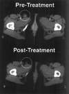
CT scan of right inguinal lymph node before and after gene transfer and TIL adoptive transfer. Serial sections of the pelvic region were obtained, and lymph node size was evaluated before adoptive transfer (Upper) and 9 months after transfer (Lower). A responsive right inguinal lymph node is circled.

Similar articles

Cited by

References

    1. Nabel G J, Nabel E G, Yang Z, Fox B A, Plautz G E, Gao X, Huang L, Shu S, Gordon D, Chang A E. Cold Spring Harbor Symp Quant Biol. 1994;49:699–707. - PubMed
    1. Plautz G E, Yang Z, Wu B, Gao X, Huang L, Nabel G J. Proc Natl Acad Sci USA. 1993;90:4645–4649. - PMC - PubMed
    1. Felgner J H, Kumar R, Sridhar C N, Wheeler C J, Tsai Y J, Border R, Ramsey P, Martin M, Felgner P L. J Biol Chem. 1994;269:2550–2561. - PubMed
    1. San H, Yang Z, Pompili V J, Jaffe M L, Plautz G E, Xu L, Felgner J H, Wheeler C J, Felgner P, Gao X, Huang L, Gordon D, Nabel G J, Nabel E G. Hum Gene Ther. 1993;4:781–788. - PubMed
    1. Nabel G J, Chang A E, Nabel E G, Plautz G E, Ensminger W, Fox B A, Felgner P, Shu S, Cho K. Hum Gene Ther. 1994;5:57–77. - PubMed

Publication types

MeSH terms