. 1996 Dec 24;93(26):15423-8.
doi: 10.1073/pnas.93.26.15423.

Nitric oxide-related species inhibit evoked neurotransmission but enhance spontaneous miniature synaptic currents in central neuronal cultures

Affiliations

Nitric oxide-related species inhibit evoked neurotransmission but enhance spontaneous miniature synaptic currents in central neuronal cultures

Z H Pan et al. Proc Natl Acad Sci U S A. .

Abstract

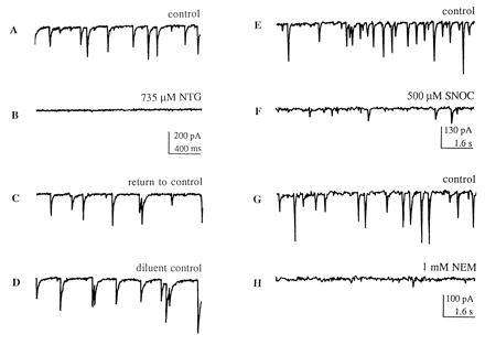
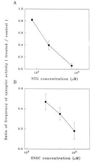
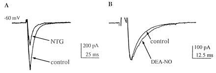
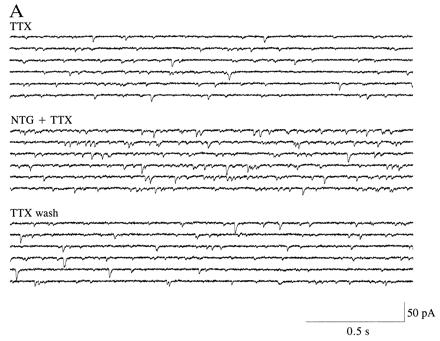
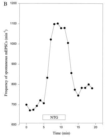
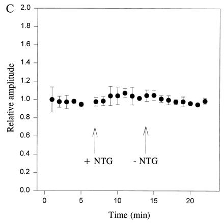
Nitric oxide (NO.) does not react significantly with thiol groups under physiological conditions, whereas a variety of endogenous NO donor molecules facilitate rapid transfer to thiol of nitrosonium ion (NO+, with one less electron than NO.). Here, nitrosonium donors are shown to decrease the efficacy of evoked neurotransmission while increasing the frequency of spontaneous miniature excitatory postsynaptic currents (mEPSCs). In contrast, pure NO donors have little effect (displaying at most only a slight increase) on the amplitude of evoked EPSCs and frequency of spontaneous mEPSCs in our preparations. These findings may help explain heretofore paradoxical observations that the NO moiety can either increase, decrease, or have no net effect on synaptic activity in various preparations.


Figures

Figure 1
Figure 1
Inhibitory effect of NO donor compounds and NEM on synaptic activity in “mass cultures.” (AC) In a representative cortical neuron, 735 μM NTG reversibly blocked synaptic transmission evoked by endogenous activity in the network of neuronal contacts (recording obtained immediately after washout of NTG); 200 μM NTG also resulted in significant suppression of synaptic activity (Fig. 2A). Holding potential, −60 mV. (D) A control solution, consisting of the diluent, propylene glycol and ethanol, plus glycerol (the 3-carbon backbone to which three -ONO2 groups are attached to form NTG) produced no effect. (E and F) A second NO group donor, SNOC, also reversibly decreased the frequency of synaptic activity in a second cortical neuron. (G and H) NEM (1 mM) mimicked the inhibitory effect of nitroso-compounds by blocking synaptic activity; this inhibitory effect was irreversible. Similar results to those illustrated in this figure were obtained in at least 8 cortical neurons from multiple platings of mass cultures.
Figure 2
Figure 2
NO+ donor compounds inhibit synaptic activity of mass cultures in a dose-dependent manner. (A) Dose-dependent inhibitory effect of NTG on the frequency of postsynaptic currents, normalized to control conditions. Data points represent means ± SEM (for n = 15 cortical neurons in mass culture). (B) Dose-dependent inhibitory effect of SNOC on the frequency of postsynaptic currents (for n = 11 neurons).
Figure 3
Figure 3
Synaptic currents elicited by depolarizing pulses at autapses in micro-cultures are decreased by NO donors of NO+ character, like NTG, but not by NO donors releasing nitric oxide (NO·), such as DEA/NO. (A) Compared with control (lower trace), NTG (500 μM) inhibited autaptic non-NMDA EPSCs on cultured, single hippocampal neurons. The EPSC amplitude recovered within 2 min of NTG washout. (B) Compared with control (upper trace), DEA/NO (1000 μM) did not inhibit the autaptic EPSC and, if anything, resulted in a minor prolongation of the synaptic current. Similar results were obtained in six autapses in micro-cultures.
Figure 4
Figure 4
Frequency of spontaneous mEPSCs is increased at autapses in micro-cultures by an NO donor with NO+ character. (A) Spontaneous mEPSCs recorded in the presence of TTX (1 μM) before and after the addition of NTG (1000 μM). The control solution superfused prior and subsequent to NTG also contained the diluent plus glycerol, as described in Fig. 1D. The frequency of mEPSCs was noticeably enhanced in the presence of NTG. (B) Plot of frequency of spontaneous mEPSCs versus time for a hippocampal autapse. TTX (1 μM) was added to the bathing solution beginning 40 min before the epoch of recording illustrated here. NTG (1000 μM) increased the frequency of mEPSCs. Data are shown from one representative recording of 10. (C) Plot of relative amplitude of spontaneous mEPSCs versus time for hippocampal autapses. The amplitude of mEPSCs was unaffected by NTG (1000 μM), nor was there a change in the noise level that would have affected the detectability of mEPSCs. Data represent the mean ± SEM for four neurons in micro-cultures treated in an identical fashion.
Figure 4
Figure 4
Frequency of spontaneous mEPSCs is increased at autapses in micro-cultures by an NO donor with NO+ character. (A) Spontaneous mEPSCs recorded in the presence of TTX (1 μM) before and after the addition of NTG (1000 μM). The control solution superfused prior and subsequent to NTG also contained the diluent plus glycerol, as described in Fig. 1D. The frequency of mEPSCs was noticeably enhanced in the presence of NTG. (B) Plot of frequency of spontaneous mEPSCs versus time for a hippocampal autapse. TTX (1 μM) was added to the bathing solution beginning 40 min before the epoch of recording illustrated here. NTG (1000 μM) increased the frequency of mEPSCs. Data are shown from one representative recording of 10. (C) Plot of relative amplitude of spontaneous mEPSCs versus time for hippocampal autapses. The amplitude of mEPSCs was unaffected by NTG (1000 μM), nor was there a change in the noise level that would have affected the detectability of mEPSCs. Data represent the mean ± SEM for four neurons in micro-cultures treated in an identical fashion.

References

    1. Böhme G A, Bon C, Stutzmann J M, Doble A, Blanchard J C. Eur J Pharmacol. 1991;199:379–381. - PubMed
    1. Schuman E M, Madison D V. Science. 1991;254:1503–1506. - PubMed
    1. O’Dell T J, Hawkins R D, Kandel E R, Arancio O. Proc Natl Acad Sci USA. 1991;88:11285–11289. - PMC - PubMed
    1. Haley J E, Wilcox G L, Chapman P F. Neuron. 1992;8:211–216. - PubMed
    1. Gribkoff V K, Lum-Ragan J T. J Neurophysiol. 1992;68:639–642. - PubMed

Publication types

MeSH terms

LinkOut - more resources