. 1996 Dec 24;93(26):15469-74.
doi: 10.1073/pnas.93.26.15469.

Regeneration of broken tip links and restoration of mechanical transduction in hair cells

Affiliations

Regeneration of broken tip links and restoration of mechanical transduction in hair cells

Y Zhao et al. Proc Natl Acad Sci U S A. .

Abstract

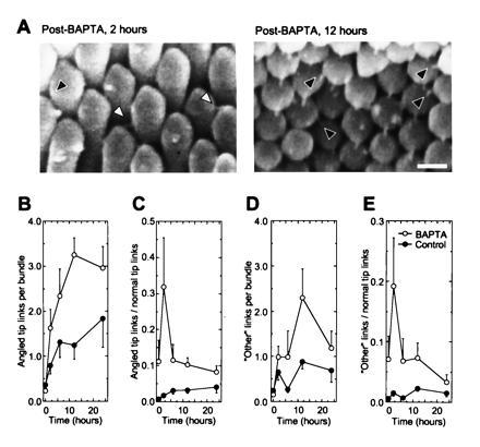
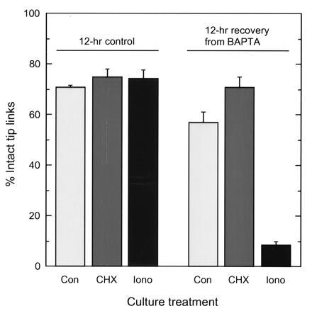
A hair cell's tip links are thought to gate mechanoelectrical transduction channels. The susceptibility of tip links to acoustic trauma raises questions as to whether these fragile structures can be regenerated. We broke tip links with the calcium chelator 1,2-bis(O-aminophenoxy) ethane-N,N,N',N'-tetraacetic acid and found that they can regenerate, albeit imperfectly, over several hours. The time course of tip-link regeneration suggests that this process may underlie recovery from temporary threshold shifts induced by noise exposure. Cycloheximide does not block tip-link regeneration, indicating that new protein synthesis is not required. The calcium ionophore ionomycin prevents regeneration, suggesting regeneration normally may be stimulated by the reduction in stereociliary Ca2+ when gating springs rupture and transduction channels close. Supporting the equivalence of tip links with gating springs, mechanoelectrical transduction returns over the same time period as tip links; strikingly, adaptation is substantially reduced, even 24 hr after breaking tip links.


Figures

Figure 1
Figure 1
Tip links regenerate after BAPTA treatment. Tip links are defined by their electron-microscopic appearance along a hair bundle’s axis of mirror symmetry, the 1,00 lattice plane of Tilney and coworkers (9). (A) Scanning electron microscopic view of individual hair bundles before or after treatment with BAPTA. Bundles from basilar papillae cultured for 24 hr after BAPTA treatment have tip links in equal numbers to bundles from control papillae. (Bar = 1 μm.) (B) Compilation of data from 162 bundles. BAPTA-treatment data were fit with the equation {% of intact tip links = A + B[1 − exp(−t/τ)]n}, where A is the fraction of intact tip links after BAPTA treatment (4%) and B is the increase in tip links during regeneration (60%). With the time constant τ as the only unconstrained variable, values for τ were as follows: for n = 1, τ = 6.6 hr (r = 0.98); for n = 2, τ = 3.9 hr (r = 0.997); and for n = 3, τ = 3.1 hr (r = 0.998). The fit for n = 2 is plotted here. Control data were plotted with an exponential fit with a time constant of about 185 hr.
Figure 2
Figure 2
Ultrastructure of control and regenerated tip links. Basilar papillae were treated with BAPTA or a control solution, and then fixed and processed for transmission electron microscopy immediately or after a culture period of 12 hr. Tips links in control papillae appeared as previously described (for examples, see refs. , , , and 11); they occasionally appeared to be branched and were often associated with amorphous material, possibly a glycocalyx. If present, osmiophilic insertional densities at the tip of a stereocilium and on the side of its neighbor clearly marked tip-link ends (densities are most clear in the hair cell that was BAPTA-treated, but not cultured). Dimensions and features of regenerated tip links fell into ranges seen in control papillae. In no case did we see osmiophilic densities at ends of regenerated tip links that were as evident as those seen in control papillae. (Bar = 100 nm.)
Figure 3
Figure 3
Angled tip links increase in frequency after tip-link regeneration. (A) Angled tip links (black arrowheads) and “other” links (white arrowheads). Angled tip links are linkages seen in scanning electron micrographs that have the same apparent diameter and length as normal tip links but connect a short stereocilium to a taller neighbor immediately adjacent to the closest stereocilium aligned along axis of mirror symmetry; because of hexagonal packing of stereocilia, these links are found at 60° to the axis of mirror symmetry (the 1,0 lattice plane of ref. 9). “Other” links also have similar apparent length and diameter but are found below normal tip link positions. “Other” links were relatively rare in normal bundles. (Bar = 250 nm.) (BE) Absolute and relative numbers of angled tip links and “other” links during basilar-papilla culture. Two hours after BAPTA treatment, the relative numbers of angled tip links and “other” links increases sharply. Each observer counted angled tip links on the same 162 bundles as in Fig. 1B. For each bundle, the four observers’ estimates were averaged; at each time point, means ± standard errors derived from 15–20 bundles are reported.
Figure 4
Figure 4
Effects of cycloheximide and ionomycin on tip-link regeneration. Basilar papillae were treated with BAPTA or a control solution and then incubated for 12 hr with no treatment (Con), 40 μM cycloheximide (CHX), or 1 μM ionomycin (Iono). For each experimental condition, means ± SEs derived from 14–22 bundles acquired from 6–12 papillae are reported.
Figure 5
Figure 5
Mechanoelectrical transduction returns after BAPTA treatment. (A) Control hair cell. (Inset) Disruption of mechanoelectrical transduction (single displacements) by bath application of 5 mM BAPTA on an isolated hair cell. (B) Hair cell isolated 1 hr after 15-min treatment of the basilar papilla with 5 mM BAPTA. Transduction currents are absent. (C) Hair cell isolated from control basilar papilla cultured for 24 hr. Although transduction-current amplitudes and adaptation rates were unaffected by 24 hr culture, the extent of adaptation was diminished. (D) Hair cell isolated from BAPTA-treated basilar papilla, cultured for 24 hr. Transduction-current amplitudes resembled those from control cells, but rate constants for adaptation to positive displacements were diminished by more than 5-fold. (E) Instantaneous displacement-response relationships (for different cells than those in AD).
Figure 6
Figure 6
Proposed model for tip-link regeneration. (A) After rupture of tip links, the concentration of Ca2+ drops in stereocilia, and tip-link regeneration is favored. In this diagram, two channels, one with an attached tip-link subunit, have been brought into the putative “assembly zone” by molecular motors. These motors have moved from the base of each stereocilium toward its tip, with the direction of movement indicated by arrows. The motor isozyme responsible for tip-link regeneration could be the same as that making up the mature adaptation-motor assembly, or it could be a distinct isozyme. If the two motors bring into close proximity a tip link and its receptor, such as a transduction channel, the tip link can bind to its receptor. The motors then move the transduction apparatus to its final resting position. The depiction of the tip link as being initially bound to the shorter stereocilium is arbitrary; in addition, although channels are shown at both tip-link ends, they may be randomly distributed between upper and lower ends (12). (B) After tip links have been regenerated, functional transduction channels maintain a modestly elevated Ca2+ concentration close to stereociliary tips (16). In our model, this elevated Ca2+ concentration prevents assembly of additional tip links. Over a time period that is presumably longer than 24 hr, the adaptation motor matures to give fast kinetics.

References

    1. Hudspeth A J. Nature (London) 1989;341:397–404. - PubMed
    1. Pickles J O, Corey D P. Trends Neurosci. 1992;15:254–259. - PubMed
    1. Corey D P, Hudspeth A J. J Neurosci. 1983;3:962–976. - PMC - PubMed
    1. Pickles J O, Comis S D, Osborne M P. Hear Res. 1984;15:103–112. - PubMed
    1. Assad J A, Shepherd G M G, Corey D P. Neuron. 1991;7:985–994. - PubMed

Publication types

Substances

LinkOut - more resources