. 1997 Jan 1;17(1):171-80.
doi: 10.1523/JNEUROSCI.17-01-00171.1997.

Specialized membrane domains for water transport in glial cells: high-resolution immunogold cytochemistry of aquaporin-4 in rat brain

Affiliations

Specialized membrane domains for water transport in glial cells: high-resolution immunogold cytochemistry of aquaporin-4 in rat brain

S Nielsen et al. J Neurosci. .

Abstract

Membrane water transport is critically involved in brain volume homeostasis and in the pathogenesis of brain edema. The cDNA encoding aquaporin-4 (AQP4) water channel protein was recently isolated from rat brain. We used immunocytochemistry and high-resolution immunogold electron microscopy to identify the cells and membrane domains that mediate water flux through AQP4. The AQP4 protein is abundant in glial cells bordering the subarachnoidal space, ventricles, and blood vessels. AQP4 is also abundant in osmosensory areas, including the supraoptic nucleus and subfornical organ. Immunogold analysis demonstrated that AQP4 is restricted to glial membranes and to subpopulations of ependymal cells. AQP4 is particularly strongly expressed in glial membranes that are in direct contact with capillaries and pia. The highly polarized AQP4 expression indicates that these cells are equipped with specific membrane domains that are specialized for water transport, thereby mediating the flow of water between glial cells and the cavities filled with CSF and the intravascular space.


Figures

Fig. 1.
Fig. 1.
AQP4 immunoblots of membrane fractions from rat brain. A, Membrane fraction (10 μg/lane) of cerebellum (Cer), spinal cord (spin), cerebral cortex (brc), thalamus (thal; and parts of hypothalamus), and medulla oblongata (m.obl). The immunoblot was reacted with affinity-purified anti-AQP4 (LL182AP). B, Identical to A except that the blot was reacted with affinity-purified antibody previously reacted overnight with immunizing peptide LL182. C, Immunoblot of membrane fractions (10 μg/lane) reacted with the second affinity-purified anti-AQP4 antibody (LL179AP). D, Immunoblot of membrane fractions from rat cerebellum using 4000 × g(4), 17,000 × g(17), and 200,000 × g(200) membrane pellets. The blot was probed with anti-AQP4 (LL182). Note that longer exposures to radiographic film of LL182 and LL179 blots revealed the presence of higher-molecular-weight bands in all lanes that contained the predominant ∼30 kDa band (not shown).
Fig. 2.
Fig. 2.
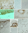
Immunocytochemical localization of AQP4 in rat brain. A, Cryosection of cerebellar cortex.Arrowheads and arrows indicate labeled glial processes in contact with granule cells and Purkinje cells. Magnification, 1000×. B, Control using affinity-purified antibody preabsorbed with excess immunizing peptide reveals no labeling. Magnification, 480×. C, Cryosection of cerebellum incubated with anti-AQP3 (Ecelbarger et al., 1995) demonstrates no labeling. Magnification, 480×. D, Vibratome section of the ventral brain surface at the level of the mesencephalon. Labeling is concentrated close to intracerebral vessels (arrows) and pia but is not associated with vessels in the subarachnoidal space (asterisk) or with arachnoid trabeculae. Magnification, 270×. E, Cryosection of thalamus demonstrates the predominant labeling of glial processes in the vicinity of vessels (arrowheads). Endothelial cells (arrows) and neurons are unlabeled. F, Cryosection of subfornical organ. Perivascular glial processes are heavily labeled (arrowheads). Distinct labeling of basolateral plasma membranes of ependymal cells is also present (arrows). Magnification, 480×. G, Higher magnification of basolateral labeling of ependymal cells (arrows) covering the subfornical organ and heavy labeling of perivascular glial cells (arrowhead). Magnification, 1000×. H, Immunolabeling control. Magnification, 480×. I, Cryosection from spinal cord reveals strong labeling of glial cells (arrowheads) and perivascular glial processes (arrows). Magnification, 480×.
Fig. 3.
Fig. 3.
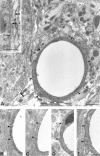
AQP4 in glial processes. Low-magnification electron micrograph showing the distribution of AQP4 immunoreactivity in the Purkinje cell layer (A) and granule cell layer (B) of the cerebellum. Immunogold particles are present along perivascular glial processes (arrowheads) and on glial processes apposed to the Purkinje cell (double arrowheads). The frame in Aindicates area enlarged in inset. Immunolabeling is abolished by preadsorption with the peptide used for immunization (C) and by substituting nonimmune IgG for the primary antibody (D); immunolabeling is not reduced after adsorption with a PKC-γ peptide of same length as the immunizing AQP4 peptide (E). Bax, Basket cell axon;P, Purkinje cell. Scale bars: A, 1 μm;E, inset in A, 0.5 μm.BD are the same magnification asE.
Fig. 4.
Fig. 4.
Polarized expression and membrane topology of AQP4 in glial cells. Many immunogold particles are present along glial membranes facing blood vessels (A, B,D, E) and pia (C), but few particles overlie membranes facing the neuropil (double-headed arrows indicate the two membranes). B, After preembedding immunogold labeling, silver-intensified immunogold particles localize AQP4 at the cytoplasmic face of the membrane (compare postembedding labeling in A andD). D, Double labeling with antibodies to AQP4 (30 nm gold particles) and the glutamate transporter GLAST (15 nm) reveals distribution of the two antigens to membranes at the opposite poles of the cell. Unlike AQP4, GLAST is concentrated along glial membranes apposed to the neuropil, including those that contact parallel fiber (Pf) synapses with Purkinje cell spines (S). E, Postembedding immunogold labeling of cryosection confirms selective labeling of the perivascular glial membrane. End, Endothelium; Gr, granule cell; Pf, parallel fiber terminal;S, Purkinje cell spines; large asterisk, pial surface; small asterisks, endothelial basal lamina;arrowheads, glial lamellae apposed to parallel fiber synapses. Scale bars: AD, 0.5 μm;E, 1 μm.
Fig. 5.
Fig. 5.
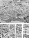
AQP4 in glial lamellae but not in neurons of the supraoptic nucleus. A, The ventral glial lamina (arrowhead in inset) associated with the supraoptic nucleus contains numerous glial lamellae that are heavily decorated with immunogold particles. The fibroblast (Fi) in the subarachnoidal space (asterisk) is unlabeled.Co, Collagen fibers; Gf, glial filaments.A, Inset, Sagittal vibratome section 1.9 mm lateral to midline after light immunolabeling for AQP4. The predominant staining lies along the ventral brain surface corresponding to the position of the supraoptic nucleus (including its retrochiasmatic portion) and in the cerebellum (Cb). Hi, Hippocampus;Ot, optic tract; Th, thalamus.BD, Immunogold particles identify AQP4 in glial membranes associated with pia (double-headed arrow in C), blood vessels (double-headed arrow in D), and magnocellular neurons (double arrowheads in B), but not in neuronal membranes (arrow in inset).End, Endothelial cell; Gf, glial filaments; MN, magnocellular neuron;asterisk, pial surface. Frame inB shows area enlarged in inset. Scale bars: B, 1 μm; A, C,D, inset in B, 0.5 μm;inset in A, 5 mm.
Fig. 6.
Fig. 6.
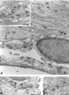
Glial and ependymal expression of AQP4 in the subfornical organ. A, Immunogold particles identify AQP4 along the entire glial lamellae except at the membrane domains engaged in gap or adhaerens type junctions (arrows) or contacting neuronal elements (double-headed arrow ininset). The vessel (V) and associated basal laminae (asterisks) are devoid of AQP4 immunolabeling. Co, Collagen; Fi, fibroblast; Gf, glial filaments; PVS, perivascular space. A, Inset, Unlabeled synapses (arrowheads) sandwiched between glial lamellae. The adjacent glial processes are polarized with respect to AQP4 expression (double-headed arrow). De, Dendrite. B, AQP4 labeling of lateral but not apical membranes of ependymal cells covering the subfornical organ (compare Fig. 2). Immunogold particles (double arrowheads) lie between junctional membrane specializations. C, Choroid plexus cells and microvilli (Mi) do not express AQP4.Arrow, Apical junctional complex. Scale bars:A, C, 1 μm; B,inset in A, 0.5 μm.

Similar articles

Cited by

References

    1. Agre P, Preston GM, Smith BL, Jung JS, Raina S, Moon C, Guggino WB, Nielsen S. Aquaporin CHIP: the archetypal molecular water channel. Am J Physiol. 1993;265:F463–F476. - PubMed
    1. Armstrong WE, Schoeler J, McNeill TH. Immunocytochemical, Golgi and electron microscopic characterization of putative dendrites in the ventral glial lamina of the rat supraoptic nucleus. Neuroscience. 1982;7:679–694. - PubMed
    1. Chaudhry FA, Lehre KP, van Lookeren Campagne M, Ottersen OP, Danbolt NC, Storm-Mathisen J. Glutamate transporters in glial plasma membranes: highly differentiated localizations revealed by quantitative ultrastructural immunocytochemistry. Neuron. 1995;15:711–720. - PubMed
    1. Ecelbarger C, Terris J, Frindt G, Echevarria M, Marples D, Nielsen S, Knepper MA. Aquaporin-3 water channel localization and regulation in rat kidney. Am J Physiol. 1995;269:F663–F672. - PubMed
    1. Fitzsimons JT. Physiology and pathophysiology of thirst and sodium appetite. In: Seldin DW, Giebisch G, editors. The kidney: physiology and pathophysiology. Raven; New York: 1992. pp. 1615–1648.

Publication types

MeSH terms

LinkOut - more resources