. 1997 Jan 1;17(1):241-50.
doi: 10.1523/JNEUROSCI.17-01-00241.1997.

Actin plays a role in both changes in cell shape and gene-expression associated with Schwann cell myelination

Affiliations

Actin plays a role in both changes in cell shape and gene-expression associated with Schwann cell myelination

C Fernandez-Valle et al. J Neurosci. .

Abstract

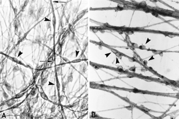
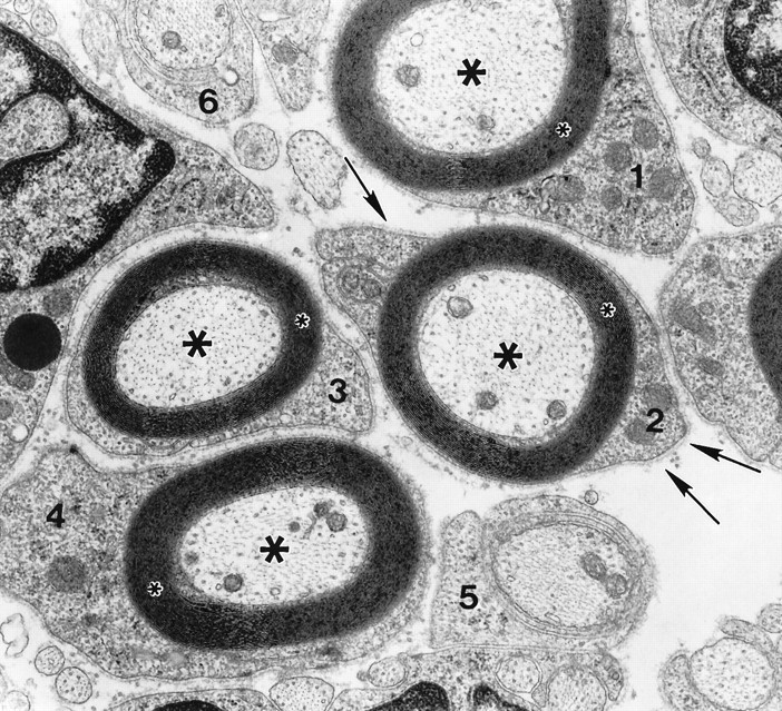
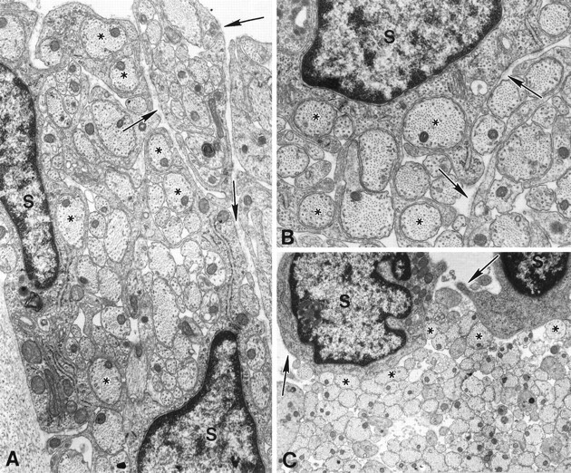
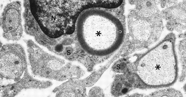
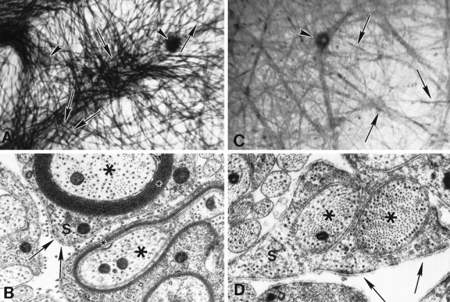
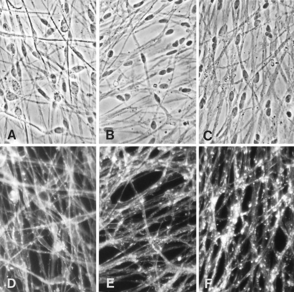
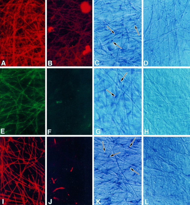
Schwann cell (SC) differentiation into a myelinating cell requires concurrent interactions with basal lamina and an axon destined for myelination. As SCs differentiate, they undergo progressive morphological changes and initiate myelin-specific gene expression. We find that disrupting actin polymerization with cytochalasin D (CD) inhibits myelination of SC/neuron co-cultures. Basal lamina is present, neurons are healthy, and the inhibition is reversible. Electron microscopic analysis reveals that actin plays a role at two stages of SC differentiation. At 0.75-1.0 microg/ml CD, SCs do not differentiate and appear as "rounded" cells in contact with axons. This morphology is consistent with disruption of actin filaments and cell shape changes. However, at 0.25 microg/ml CD, SCs partially differentiate; they elongate and segregate axons but generally fail to form one-to-one relationships and spiral around the axon. In situ hybridizations reveal that SCs in CD-treated cultures do not express mRNAs encoding the myelin-specific proteins 2',3'-cyclic nucleotide phosphodiesterase (CNP), myelin-associated glycoprotein (MAG), and P0. Our results suggest that at the lower CD dose, SCs commence differentiation as evidenced by changes in cell shape but are unable to elaborate myelin lamellae because of a lack of myelin-specific mRNAs. We propose that F-actin influences myelin-specific gene expression in SCs.


Figures

Fig. 1.
Fig. 1.
CD inhibits Schwann cell differentiation on collagen. SC–sensory neurons cultures were grown for 1 week in myelination-permissive medium alone (A) or with 1 μg/ml CD (B). Sudan black staining used to visualize myelin revealed that SCs (arrowheads) in control cultures (A) had flattened and elongated along axons and were beginning to form myelin (between arrows). SCs grown in the presence of CD (B) failed to elongate in association with axons, retained a rounded morphology, and did not myelinate axons. Magnification, 800×.
Fig. 2.
Fig. 2.
Schwann cells cultured in myelination-permissive medium rapidly differentiate to myelin-forming cells. This electron micrograph reveals the degree of SC differentiation achieved during 1 week in culture. Four SCs (1–4) elaborated up to 25 myelin lamellae (small asterisks) around axons (large asterisks). Each SC–axon unit is surrounded by a complete basal lamina (arrows). Two SCs (5, 6) at earlier stages of differentiation have begun to spiral around smaller diameter axons. Magnification, 30,000×.
Fig. 3.
Fig. 3.
CD causes a dose-dependent disruption of morphological differentiation; elongation and spiralization are selectively inhibited by different CD concentrations. SCs were cultured in myelination-permissive medium containing 0.5 (A), 0.75 (B), or 1.0 μg/ml (C) CD for 1 week. At the lowest CD concentration (A), SCs (S) elongated and extended multiple processes into axonal fascicles, thereby segregating axons into smaller groups or isolating them completely. Continuing spiralization around an individual axon was not observed. At the intermediate CD concentration (B), SCs retained the ability to extend processes and segregate axons into smaller groups. However, at the highest CD concentration (C), SCs remained rounded, adhered to axons (asterisks) but did not extend processes into axonal fascicles. Basal lamina (arrows) assembled and attached to the SC surface in all CD doses. Magnification:A, 25,000×, B, 35,000×,C, 15,000×.
Fig. 5.
Fig. 5.
CD’s inhibitory effect on myelination is reversible. Cultures were grown in myelination-permissive medium with 0.75 μg/ml CD for 1 week and then without CD for an additional week to determine whether drug toxicity prevented SC function. At removal of CD, SCs spiraled membranes around axons. An SC (S), shown with its nucleus (N), elaborated 20 myelin lamellae (small asterisk) around an axon (large asterisks), and an adjacent SC (S) is beginning to myelinate. Magnification, 25,000×.
Fig. 4.
Fig. 4.
Lower CD concentrations inhibit myelin formation in cultures grown on laminin instead of collagen. Cultures grown in myelination-permissive medium alone (A,B) or with CD, 0.25 μg/ml (C,D) are illustrated. Sudan black staining (A, C) demonstrates that myelin (arrows) is abundant in control cultures but is generally absent in CD-treated cultures, although SCs elongate along axons. Neuron cell bodies are indicated by arrowheads. EM confirms that in myelination-permissive medium (B), SCs (S) differentiate and form myelin (small asterisks). In CD-treated cultures (D), myelin is absent, but basal lamina is present (arrows). SCs retain the ability to segregate axons (large asterisks) but cannot spiral membrane around the axon to form myelin lamellae in 0.25 μg/ml CD. Magnification: A, C, 166×; B, D, 40,000×.
Fig. 6.
Fig. 6.
Actin organization is progressively disrupted with increasing CD concentrations. Phase (AC) and fluorescent (DF) micrographs of cultures grown on laminin for 1 week in myelination-permissive medium alone (A, D) or with 0.25 μg/ml CD (B, E), or 0.5 μg/ml CD (C, F). Cultures were fixed and stained with rhodamine-conjugated phalloidin to visualize the distribution of actin filaments. In the absence of CD, phalloidin staining appears continuous and linear. In the presence of CD, the pattern of staining becomes disrupted, appearing as large fluorescent aggregates. Magnification, 500×.
Fig. 7.
Fig. 7.
CD inhibits SC expression of myelin-specific proteins and mRNAs. Cultures grown in myelination-permissive medium alone (A, C, E,G, I, K) or with 0.25 μg/ml CD (B, D, F,H, J, L) were immunostained with antibodies against CNP (A,B), MAG (E, F), or P0 (I, J) or processed forin situ hybridization using CNP (C,D), MAG (G, H), or P0 RNA probes (K, L) to detect myelin-specific mRNAs. Arrows indicate positively stained Schwann cells. CNP, MAG, and P0 expression was greatly reduced in SCs cultured in the CD. In situ hybridization results indicated that mRNAs encoding the myelin-specific proteins were either not expressed or expressed at very low levels compared with control cultures. Sister cultures were hybridized with sense probes for each mRNA as negative controls. Magnification, 600×.

Similar articles

Cited by

References

    1. Aguayo AJ, Charron L, Bray GM. Potential of Schwann cells from unmyelinated nerves to produce myelin: a quantitative ultrastructural and radiographic study. J Neurocytol. 1976a;5:565–573. - PubMed
    1. Aguayo AJ, Epps J, Charron L, Bray GM. Multipotentiality of Schwann cells in cross-anastomosed and grafted myelinated and unmyelinated nerves: quantitative microscopy and radioautography. Brain Res. 1976b;104:1–20. - PubMed
    1. Barth J, Ivarie R. Polyvinyl alcohol enhances detection of low abundance transcripts in early stage quail embryos in a nonradioactive whole mount in situ hybridization technique. Biotechniques. 1994;17:324–326. - PubMed
    1. Bermingham JR, Scherer SS, O’Connell S, Arroyo E, Kalla KA, Powell FL, Rosenfeld MG. Tst-1/Oct-6/SCIP regulates a unique step in peripheral myelination and is required for normal respiration. Genes Dev. 1996;10:1751–1762. - PubMed
    1. Bolin LM, Shooter EM. Neurons regulate Schwann cell genes by diffusible molecules. J Cell Biol. 1993;123:237–243. - PMC - PubMed

Publication types

MeSH terms