. 1997 Jan 1;17(1):308-24.
doi: 10.1523/JNEUROSCI.17-01-00308.1997.

Synaptic integration of functionally diverse pallidal information in the entopeduncular nucleus and subthalamic nucleus in the rat

Affiliations

Synaptic integration of functionally diverse pallidal information in the entopeduncular nucleus and subthalamic nucleus in the rat

M D Bevan et al. J Neurosci. .

Abstract

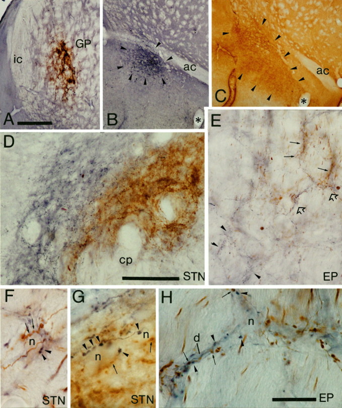
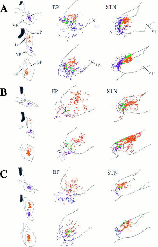
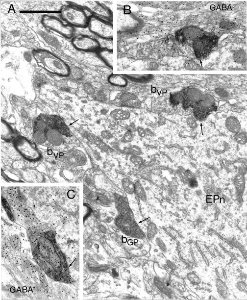
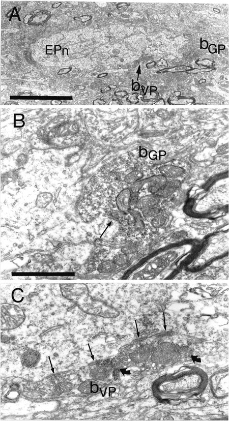
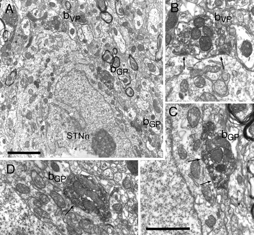
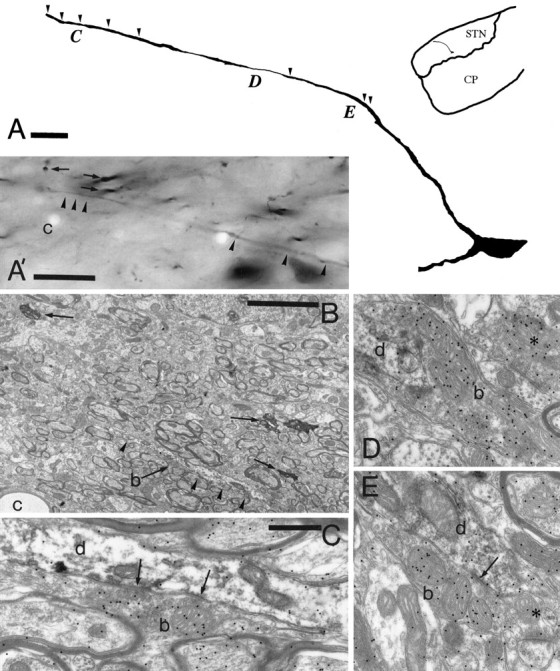
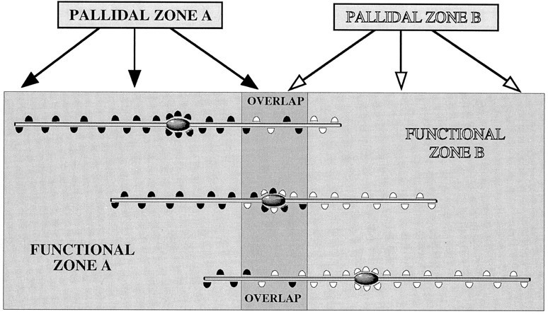
To determine the principles of synaptic innervation of neurons in the entopeduncular nucleus and subthalamic nucleus by neurons of functionally distinct regions of the pallidal complex, double anterograde labeling was carried out at both light and electron microscopic levels in the rat. Deposits of the anterograde tracers Phaseolus vulgaris-leucoagglutinin and biotinylated dextran amine were placed in different functional domains of the pallidal complex in the same animals. The tracer deposits in the ventral pallidum and the globus pallidus gave rise to GABA-immunopositive projections to the entopeduncular nucleus, the subthalamic nucleus, and the more medial lateral hypothalamus that were largely segregated but overlapped at the interface between the two fields of projection. In these regions the proximal parts of individual neurons in the entopeduncular nucleus, lateral hypothalamus, and subthalamic nucleus received synaptic input from terminals derived from both the ventral pallidum and the globus pallidus. Furthermore, the analysis of the afferent synaptic input to the dendrites of neurons in the subthalamic nucleus that cross functional boundaries of the nucleus defined by the pallidal inputs, revealed that terminals with the morphological and neurochemical characteristics of those derived from the pallidal complex make synaptic contact with all parts of the dendritic tree, including distal regions. It is concluded that functionally diverse information carried by the descending projections of the pallidal complex is synaptically integrated by neurons of the entopeduncular nucleus, lateral hypothalamus, and subthalamic nucleus by two mechanisms. First, neurons located at the interface between functionally distinct, but topographically adjacent, projections could integrate diverse information by means of the synaptic convergence at the level of the cell body and proximal dendrites. Second, because the distal dendrites of neurons in the subthalamic nucleus receive input from the pallidum, those that extend across two distinct domains of pallidal input could also provide the morphological basis of integration.


Figures

Fig. 1.
Fig. 1.
Light micrographs of anterograde labeling from the ventral pallidum and globus pallidus (GP) in the entopeduncular nucleus (EP) and subthalamic nucleus (STN). A–C, Sites of deposit of BDA in the globus pallidus (A) were revealed by using DAB as the chromogen, and PHA-L in the ventral pallidum (B) were revealed by using nickel-DAB as the chromogen for the peroxidase reaction. The extent of the tracer deposit in the ventral pallidum (arrowheads) was assessed by staining of adjacent sections to reveal substance P immunoreactivity (arrowheads) (C). D, Medium-power micrograph of anterograde labeling in the subthalamic nucleus. The fibers anterogradely labeled from the ventral pallidum (blue) occupy the medial and dorsal aspects of the STN, whereas those from the globus pallidus (brown) occupy the more lateral parts; however, the two sets of anterogradely labeled fibers are mixed at the interface between the projections. Note that the width of the fields of anterograde labeling is well within the dendritic diameter of subthalamic neurons. E, Medium-power micrograph of anterograde labeling in the entopeduncular nucleus. The fibers derived from the globus pallidus (brown; some indicated by arrows) and those derived from the ventral pallidum (blue; some indicated byarrowheads) are largely separate at this level, although there are areas of overlap of the two sets of fibers (some indicated byopen arrows and shown at higher magnification inH). F, G, High-power micrographs of unstained neuronal perikarya in the STN that are closely apposed by axonal swellings derived from both the ventral pallidum (blue; some indicated by arrowheads) and the globus pallidus (brown; some indicated byarrows). Individual perikarya are apposed by several boutons from each site. H, High-power micrograph of the region of the entopeduncular nucleus indicated by open arrows in E. An unstained neuronal perikaryon (n) and dendrite (d) are closely apposed by axonal swellings derived from both the ventral pallidum (blue; some indicated by arrowheads) and the globus pallidus (brown; some indicated byarrows). ac, Anterior commissure;cp, cerebral peduncle; ic, internal capsule. Scale bars: A–C (shown in A), 500 μm; D, E, (shown in D), 100 μm;F–H (shown in H), 20 μm.
Fig. 2.
Fig. 2.
Schematic representations of the sites of deposit of PHA-L in the ventral pallidum (VP) and BDA in the globus pallidus (GP) and the site of anterograde transport in the entopeduncular nucleus (EP) and subthalamic nucleus (STN) in three animals (A–C). Dots at the injection sites represent individual neurons that have taken up the PHA-L (blue) or the BDA (red). Theblue and red stippling of the two rostrocaudal levels of the EP and STNrepresent the anterogradely labeled fibers from the VPand GP, respectively. Although the topography of the two projections is distinct, many neurons were identified (green dots) that were apposed by boutons derived from both the VP and GP.ac, Anterior commissure; cp, cerebral peduncle; ic, internal capsule.
Fig. 3.
Fig. 3.
Retrograde labeling in the striatal complex after tracer deposits in the ventral pallidum and the globus pallidus. Low-power micrograph of the neostriatum and nucleus accumbens of the same animal as illustrated in Figure 2C. The section was incubated to reveal retrogradely transported PHA-L that was injected in the ventral pallidum and BDA that was injected in the globus pallidus. Although it is difficult to distinguish the labeling in this black and white micrograph, neurons retrogradely labeled from the globus pallidus (labeled with DAB; area indicated by arrowheads) are present only in the dorsal part of the neostriatum, whereas neurons retrogradely labeled from the ventral pallidum (labeled with Ni-DAB; area indicated by arrowheads) are present only in the most ventral aspects of the neostriatum and the nucleus accumbens. ac, Anterior commissure; lv, lateral ventricle; nac, nucleus accumbens;ns, neostriatum. Scale bar, 0.5 mm .
Fig. 4.
Fig. 4.
Synaptic convergence of terminals derived from different functional domains of the pallidal complex in the entopeduncular nucleus. A, Electron micrograph of part of a proximal dendrite of a neuron in the entopeduncular nucleus (EPn). The neuron is apposed by three anterogradely labeled boutons, each of which forms symmetrical synaptic contact with the neuron (arrows). Two of the boutons contain the BDHC peroxidase reaction product that was used to localize the PHA-L anterogradely transported from the ventral pallidum (bVP). The third bouton (bGP) contains the DAB reaction product that was used to localize the BDA anterogradely transported from the globus pallidus. Note that the BDHC reaction product that labels the terminals from the ventral pallidum has an irregular appearance and occupies only part of the labeled bouton, leaving many vesicles visible that do not have reaction product associated with them. In contrast, the DAB reaction product that labels the boutons from the globus pallidus is amorphous and occupies the whole of the labeled structure.B, A serial section of the upper of the two boutons derived from the ventral pallidum. This section was processed by the post-embedding immunogold method to reveal GABA immunoreactivity. The bouton has a high density of immunogold particles associated with it (index of GABA immunoreactivity = 3.79). C, Serial section of the bouton derived from the globus pallidus labeled by the post-embedding immunogold method to reveal GABA immunoreactivity. The bouton has a high density of immunogold particles associated with it (index of GABA immunoreactivity = 7.51). Scale bar (shown in A):A–C, 1 μm.
Fig. 5.
Fig. 5.
Synaptic convergence of terminals derived from different functional domains of the pallidal complex in the entopeduncular nucleus. A, The large proximal dendrite of a neuron in the entopeduncular nucleus (EPn) is apposed by two anterogradely labeled boutons. The bouton, shown at high power in B, contains the floccular DAB reaction product that adheres to the external surface of vesicle and mitochondrial membranes and indicates that it is derived from the globus pallidus (bGP). The bouton forms symmetrical synaptic contact (arrow) with dendrite. The other bouton contains the BDHC reaction product that is granular in appearance and is located only at restricted sites in the bouton (granules indicated bycurved arrows in C), indicating that it is derived from the ventral pallidum (bVP). It forms multiple symmetrical contacts with the dendrite (arrows). Scale bars: A, 5 μm;B, 1 μm.
Fig. 6.
Fig. 6.
Synaptic convergence of terminals derived from different functional domains of the pallidal complex in the lateral hypothalamus adjacent to the entopeduncular nucleus. A, B, Low-power micrographs at two levels of a neuron in the lateral hypothalamus (LHn) that was apposed by four anterogradely labeled terminals. Three of the terminals (bVP), the positions of which are indicated byarrows and the letters C, D, andE, are shown at higher magnification in C, D, and E, respectively. Each contains the BDHC reaction product (some granules of which are indicated by curved arrows) that was used to reveal the PHA-L transported from the ventral pallidum. The reaction product characteristically occupies only part of the labeled boutons. In each case the boutons form symmetrical synaptic contact (arrows) with the perikaryon (C, D) or the dendrite (d in E). The micrograph E is a serial section of that inB; the myelinated axon (ma) is indicated by an arrowhead in B. The fourth bouton (bGP), the position of which is indicated byF in B, is shown at high magnification inF and contains the floccular DAB reaction product that adheres to membranes and occupies the whole of the labeled structure, identifying it as arising from the globus pallidus. This bouton also makes symmetrical synaptic contact (arrow) with the perikaryon of the lateral hypothalamic neuron. Glial cells (g) and a capillary (c) are labeled for correlation between the two low-power micrographs. Scale bars: A, B (shown in B), 10 μm;C–E (shown in E), 1 μm.
Fig. 7.
Fig. 7.
Synaptic convergence of terminals derived from different functional domains of the pallidal complex in the subthalamic nucleus. A, Part of the cell body of a neuron in the subthalamic nucleus (STNn) that is apposed by three anterogradely labeled terminals (bVP, bGP) shown at higher magnification inB–D. In this animal the injections were reversed, i.e., the PHA-L was injected in and anterogradely transported from the globus pallidus, and the BDA was injected in and anterogradely transported from the ventral pallidum. One of the boutons is lightly labeled with the DAB reaction product that adheres to vesicle and mitochondrial membranes, identifying it as arising in the ventral pallidum (bVP). It is shown at higher magnification inB. The bouton forms symmetrical synaptic contacts (arrows) with the subthalamic neuron. The other two boutons, shown at high magnification in C andD, are strongly labeled with the crystalline BDHC reaction product, which has an irregular appearance. These boutons are thus derived from the globus pallidus (bGP) and form symmetrical synaptic contacts with the neuron (arrows). Note that micrograph D is a different serial section of that shown in A. Scale bars:A, 2 μm; B–D (shown inC), 1 μm.
Fig. 8.
Fig. 8.
Synaptic convergence of terminals derived from different functional domains of the pallidal complex in the subthalamic nucleus. A, Cell body of a neuron in the subthalamic nucleus (STNn) that is postsynaptic to two anterogradely labeled boutons. The bouton bVP, shown at high magnification in B, contains the crystalline BDHC reaction product that occupies only part of the bouton. The BDHC was used to reveal the PHA-L transported from the ventral pallidum. The bouton forms symmetrical synaptic contact (arrow) with the neuron. The bouton bGP, shown at higher magnification in C, is intensely labeled with the floccular DAB reaction product used to localize the BDA transported from the globus pallidus. The reaction product fills the whole of the bouton and obscures most internal structures. This bouton forms symmetrical synaptic contact with the neuron (arrow). Scale bars: A, 3 μm; B, C (shown inC), 1 μm.
Fig. 9.
Fig. 9.
Topology of pallidal terminals in synaptic contact with the distal dendrites of neurons in the subthalamic nucleus. A, Drawing of a neuron in the subthalamic nucleus (inset shows the position in theSTN) that was labeled by the retrograde transport of neurobiotin from the substantia nigra. This neuron was chosen for analysis because of the extent of the dendritic labeling that crosses functional territories as defined by pallidal inputs. Thearrowheads indicate the position at which synaptic input from pallidal terminals defined on the basis of morphology and neurochemistry were identified. Three additional pallidal inputs indicated by the location of the letters C, D, andE are shown at the electron microscopic level inC, D, and E, respectively. A′, B, and C show the correlation between the light and electron microscopic levels. The distal part of the dendrite is indicated by the arrowheads in the light micrograph (A) and in the electron micrograph (B). Three other neurobiotin-labeled structures (arrows) and a capillary (c) are indicated on both micrographs for the purpose of correlation.C, A bouton (b; also indicated in micrograph B) has the morphological features of a terminal derived from the pallidal complex, forms symmetrical synaptic contact with the dendrite (d) ∼245 μm from the perikaryon, and has a high level of GABA immunoreactivity associated with it (index of GABA immunoreactivity is 5.8 compared with GABA-negative terminals forming asymmetrical synapses in the same section). The bouton (b) in D also possesses the features of a pallidal terminal, is ∼145 μm from the perikaryon, and has an index of GABA immunoreactivity of 11.2. In E the bouton (b) forms symmetrical synaptic contact with the dendrite (arrow) ∼100 μm from the perikaryon and has an index of GABA immunoreactivity of 6.54. The asterisk inD indicates a bouton that forms symmetrical synaptic contact with a dendrite and has an index of GABA immunoreactivity of 9.49. The asterisk in E indicates a terminal that forms an asymmetrical synapse with a spine and is GABA-negative (index of GABA immunoreactivity 1.19). Scale bars:A, 20 μm; A′, 20 μm;B, 5 μm; C–E (shown inC), 0.5 μm.
Fig. 10.
Fig. 10.
Schematic summary of the somatic and dendritic modes of synaptic integration of descending, functionally distinct pallidal projections in the subthalamic nucleus revealed by double anterograde labeling and electron microscopy. The pallidal complex provides projections to the subthalamic nucleus that largely maintain the functional topography. Adjacent populations of neurons, illustrated by PALLIDAL ZONE A and PALLIDAL ZONE Band giving rise to black and white boutons, respectively, although mainly innervating separate but adjacent regions of the subthalamic nucleus, also give rise to a region of overlap. The dimensions and orientation of dendrites of a large proportion of neurons in the subthalamic nucleus are such that they extend in a mediolateral or ventrodorsal direction and thus cross the functional boundaries defined by the pallidal inputs. Integration of the descending, functionally diverse information from the pallidal complex occurs by synaptic convergence on individual neurons in the subthalamic nucleus. This is mediated by two systems: synaptic integration at the level of the soma and proximal dendrites, and synaptic integration at the level of more distal dendrites. Synaptic integration of pallidal inputs occurs at the level of the soma and proximal dendrites of neurons located in the region of overlap of the pallidal projections (middle neuron). These neurons receive synaptic input to their cell bodies and proximal dendrites from neurons located in both pallidal zones. Their distal dendrites, if oriented across the boundaries, will also receive inputs from both pallidal zones. The net weights of the two pallidal inputs to these neurons are likely to be similar, although the distal dendrites will preferentially receive inputs from one or the other of the pallidal zones. The second mode of synaptic integration occurs on the distal dendrites of neurons, the cell bodies of which are located within a functional zone defined on the basis of its pallidal input (top and bottom neurons in the diagram). A high proportion of the pallidal input to these neurons is derived from the single pallidal zone (PALLIDAL ZONE Aor B) that provides the innervation of the functional zone (FUNCTIONAL ZONE A or B) within which they reside. The dendrites that cross functional boundaries, however, receive synaptic input from the topographically adjacent pallidal zone in the region of overlap and in the adjacent functional zone of the subthalamic nucleus. In this case the pallidal input will be weighted in favor of the pallidal region that projects to the zone within which the neuron (mainly) resides, but will vary with the position of the neurons in relation to the boundaries and the number of dendrites crossing the boundary. In this system also, it is possible that individual dendrites will receive inputs from a single pallidal zone. The data on which this model is based were derived from injections of tracers into broad functional zones of the pallidal complex, i.e., the limbic division (ventral pallidum) and the motor and associative division (globus pallidus) that nevertheless give rise to adjacent projections. Topographic studies suggest that these broad functional zones are themselves subdivided into small groups of functionally related neurons that are organized into multiple parallel pathways. We predict that the same principles of organization will apply to these subdivisions of the pallidal complex. Although the present model is based mostly on findings of neurons in the subthalamic nucleus, the present work and previously published data (Bevan et al., 1996) indicate that the somatic integration in areas of overlap of pallidal projections also occurs in the entopeduncular nucleus, the lateral hypothalamus, and the substantia nigra pars reticulata. Furthermore, the synaptology of the pallidal projections and the synaptology and orientation of dendrites in these regions indicate that integration also probably occurs at the level of distal dendrites (see text for references). Although synaptic convergence of functionally diverse pallidal inputs also occurs on dopamine neurons of the substantia nigra pars compacta and the ventral tegmental area (Bevan et al., 1996), the exact rules of synaptic integration remain to be established.

References

    1. Albin RL, Young AB, Penney JB. The functional anatomy of basal ganglia disorders. Trends Neurosci. 1989;12:366–375. - PubMed
    1. Alexander GE, Crutcher MD. Functional architecture of basal ganglia circuits: neural substrates of parallel processing. Trends Neurosci. 1990;13:266–271. - PubMed
    1. Alexander GE, DeLong MR, Strick PL. Parallel organization of functionally segregated circuits linking basal ganglia and cortex. Annu Rev Neurosci. 1986;9:357–381. - PubMed
    1. Alexander GE, Crutcher MD, DeLong MR. Basal ganglia-thalamocortical circuits: parallel substrates for motor, oculomotor, “prefrontal” and “limbic” functions. In: Uylings HBM, Vaneden CG, Debruin JPC, Corner MA, Feenstra MGP, editors. Prefrontal cortex. Its structure, function and pathology. Elsevier; Amsterdam: 1990. pp. 119–146. - PubMed
    1. Aosaki T, Kimura M, Graybiel AM. Temporal and spatial characteristics of tonically active neurons of the primate’s striatum. J Neurophysiol. 1995;73:1234–1252. - PubMed

Publication types

Substances

LinkOut - more resources