

Human brain language areas identified by functional magnetic resonance imaging
- PMID: 8987760
- PMCID: PMC6793702
- DOI: 10.1523/JNEUROSCI.17-01-00353.1997
Human brain language areas identified by functional magnetic resonance imaging
Abstract
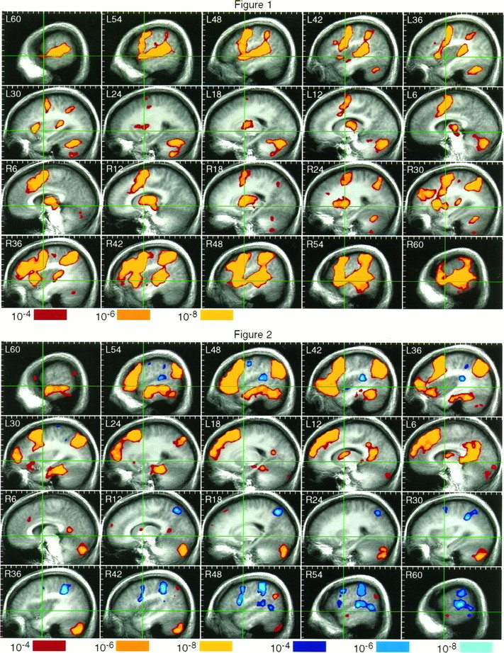
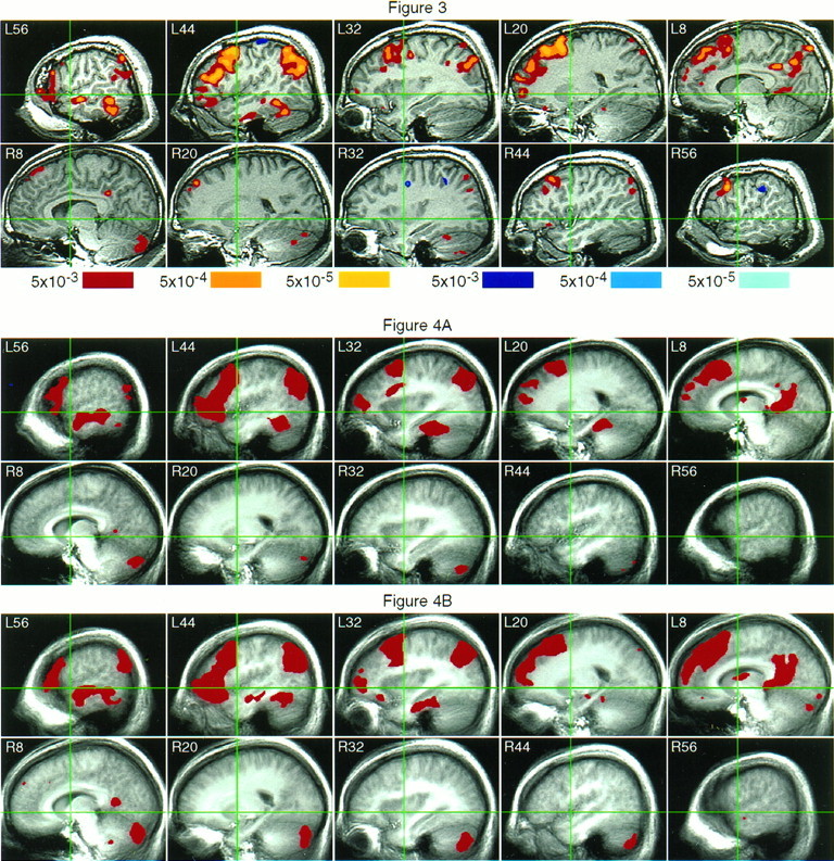
Functional magnetic resonance imaging (FMRI) was used to identify candidate language processing areas in the intact human brain. Language was defined broadly to include both phonological and lexical-semantic functions and to exclude sensory, motor, and general executive functions. The language activation task required phonetic and semantic analysis of aurally presented words and was compared with a control task involving perceptual analysis of nonlinguistic sounds. Functional maps of the entire brain were obtained from 30 right-handed subjects. These maps were averaged in standard stereotaxic space to produce a robust "average activation map" that proved reliable in a split-half analysis. As predicted from classical models of language organization based on lesion data, cortical activation associated with language processing was strongly lateralized to the left cerebral hemisphere and involved a network of regions in the frontal, temporal, and parietal lobes. Less consistent with classical models were (1) the existence of left hemisphere temporoparietal language areas outside the traditional "Wernicke area," namely, in the middle temporal, inferior temporal, fusiform, and angular gyri; (2) extensive left prefrontal language areas outside the classical "Broca area"; and (3) clear participation of these left frontal areas in a task emphasizing "receptive" language functions. Although partly in conflict with the classical model of language localization, these findings are generally compatible with reported lesion data and provide additional support for ongoing efforts to refine and extend the classical model.
Figures
References
-
- Alexander MP, Schmitt MA. The aphasia syndrome of stroke in the left anterior cerebral artery territory. Arch Neurol. 1980;37:97–100. - PubMed
-
- Alexander MP, Hiltbrunner B, Fischer RS. Distributed anatomy of transcortical sensory aphasia. Arch Neurol. 1989;46:885–892. - PubMed
-
- Barrett AM. A case of pure word-deafness with autopsy. J Nerv Ment Dis. 1910;37:73–92.
-
- Benson DF. Aphasia and related disorders: a clinical approach. In: Mesulam M-M, editor. Principles of behavioral neurology. Davis; Philadelphia: 1985. pp. 193–238.
-
- Binder JR, Rao SM, Hammeke TA, Frost JA, Bandettini PA, Hyde JS. Effects of stimulus rate on signal response during functional magnetic resonance imaging of auditory cortex. Cognit Brain Res. 1994a;2:31–38. - PubMed
Publication types
MeSH terms
Grants and funding
LinkOut - more resources
Full Text Sources
Other Literature Sources
Medical
Research Materials