. 1997 Jan 1;17(1):363-71.
doi: 10.1523/JNEUROSCI.17-01-00363.1997.

Electrophysiological and immunocytochemical evidence for a cGMP-mediated inhibition of subfornical organ neurons by nitric oxide

Affiliations

Electrophysiological and immunocytochemical evidence for a cGMP-mediated inhibition of subfornical organ neurons by nitric oxide

M Rauch et al. J Neurosci. .

Abstract

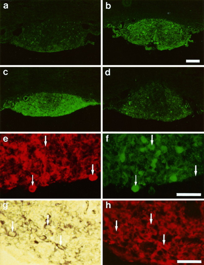
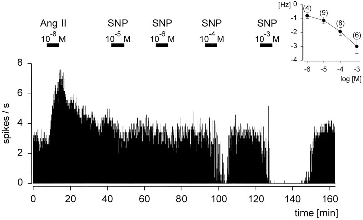
The activation of neurons in the subfornical organ (SFO) by angiotensin II (AngII) is well established and is widely regarded as the basis for the AngII-induced increase in water intake. Application of the nitric oxide (NO) donor sodium nitroprusside (SNP) led to an inhibition of the spontaneous electrical activity in 96% of the neurons sensitive for SNP (n = 50). In addition, the firing rate in 60% of the neurons inhibited by SNP decreased in response to superfusion with the natural substrate of the NO synthase (NOS) L-arginine whereas 70% increased their frequency after application of the NOS blocker NG-monomethyl-L-arginine (L-NMMA; n = 10). The inhibitory effect of SNP could be mimicked by application of membrane-permeable 8-Br-cGMP. The presence of nNOS, the neuronal isoform of NOS, was demonstrated immunocytochemically and using the NADPH-diaphorase technique on SFO slices. Using a highly selective antibody against cGMP in formaldehyde-fixed tissue, the NO donors SNP, 3-morpholinosydnonimine (SIN-1), and S-nitroso-N-acetyl-DL-penicillamine (SNAP) caused a strong increase in cGMP formation when applied under the same conditions as used for the electrophysiological recordings. These electrophysiological results suggest an important role for NO in SFO-mediated responses and offer a plausible explanation for the in vivo-observed opposite effects of AngII and NO on water intake.


Figures

Fig. 1.
Fig. 1.
Immunocytochemical and NADPH–diaphorase staining of 10-μm-thick sections of the rat subfornical organ (SFO).ad, Comparison of cGMP immunoreactivity after normal treatment (control, a) and after 10 min incubation with sodium nitroprusside (SNP; b),S-nitroso-N-acetyl-dl-penicillamine (SNAP; c), and 3-morpholinosydnonimine (SIN-1;d). Markedly increased levels of cGMP are visible after treatment with NO donors. Scale bar (shown in b): 200 μm. e, f, Double labeling of an identical SFO section for neuronal NO synthase (nNOS; e) and cGMP (f) immunoreactivity. One cell labeled by both antibodies is marked with a small arrow, whereas the large arrows mark cells exclusively stained by the cGMP antibody or nNOS antibody, indicating a codistribution rather than a colocalization. Scale bar (shown inf): 40 μm. g, h, Colocalization of NADPH–diaphorase staining (g) with nNOS immunoreactivity (h) in an identical section of the SFO. Examples of cells labeled by both techniques are marked with arrows. Scale bar (shown in h): 40 μm.
Fig. 2.
Fig. 2.
Continuous rate meter recording of a spontaneously active neuron from the rat subfornical organ showing the antagonistic effect of angiotensin II (Ang II) and sodium nitroprusside (SNP) on the same neuron. AngII and SNP were applied via superfusion during the times indicated by thehorizontal bars.
Fig. 3.
Fig. 3.
a, The inhibitory effect of sodium nitroprusside (SNP) on this neuron could be antagonized by angiotensin II (Ang II) in the presence of the NO donor. b, Repeated application of SNP was able to antagonize reversibly the excitatory effect of continuously applied AngII on this neuron.
Fig. 4.
Fig. 4.
Dose-dependent effect of sodium nitroprusside (SNP) on an angiotensin II (Ang II)-sensitive neuron, which was consecutively superfused during the times indicated by the horizontal bars. Theinset shows the averaged dose–response curve of the inhibitory effect of SNP on rat subfornical organ neurons.
Fig. 5.
Fig. 5.
Continuous rate meter recording of a spontaneously active neuron from the subfornical organ, demonstrating the effects of the natural substrate for NOS l-arginine (L-Arg), the false substrateNG-monomethyl-l-arginine (L-NMMA), and their biologically inactived-enantiomers (D-Arg,D-NMMA), which serve as controls. Thehorizontal bars represent the times of the respective superfusion.
Fig. 6.
Fig. 6.
Continuous rate meter recording of a spontaneously active neuron from the rat subfornical organ. During the times indicated by the horizontal bars, sodium nitroprusside (SNP) or the membrane-permeable analog 8-Br-cGMP (8 Br-cGMP) was applied via superfusion. 8-Br-cGMP mimics the inhibitory effect of the NO donor SNP on the same neuron.

References

    1. Calapai G, Squadrito F, Altavilla D, Zingarelli B, Campo GM, Cilia M, Caputi AP. Evidence that nitric oxide modulates drinking behaviour. Neuropharmacology. 1992;31:761–764. - PubMed
    1. Calapai G, Mazzaglia G, Cilia M, Zingarelli B, Squadrito F, Caputi AP. Mediation by nitric oxide formation in the preoptic area of endotoxin and tumour necrosis factor-induced inhibition of water intake in the rat. Br J Pharmacol. 1994;111:1328–1332. - PMC - PubMed
    1. de Vente J, Steinbusch HW. On the stimulation of soluble and particulate guanylate cyclase in the rat brain and the involvement of nitric oxide as studied by cGMP immunocytochemistry. Acta Histochem. 1992;92:13–38. - PubMed
    1. de Vente J, Schipper J, Steinbusch HW. Formaldehyde fixation of cGMP in distinct cellular pools and their recognition by different cGMP-antisera: an immunocytochemical study into the problem of serum specificity. Histochemistry. 1989;91:401–412. - PubMed
    1. Ehrlich KJ, Fitts DA. Atrial natriuretic peptide in the subfornical organ reduces drinking induced by angiotensin or in response to water deprivation. Behav Neurosci. 1991;104:365–372. - PubMed

Publication types

MeSH terms

LinkOut - more resources