

Age-related differences in neural activity during memory encoding and retrieval: a positron emission tomography study
- PMID: 8987764
- PMCID: PMC6793692
- DOI: 10.1523/JNEUROSCI.17-01-00391.1997
Age-related differences in neural activity during memory encoding and retrieval: a positron emission tomography study
Abstract
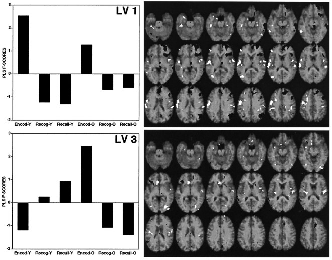
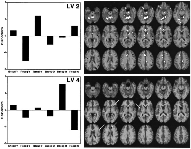
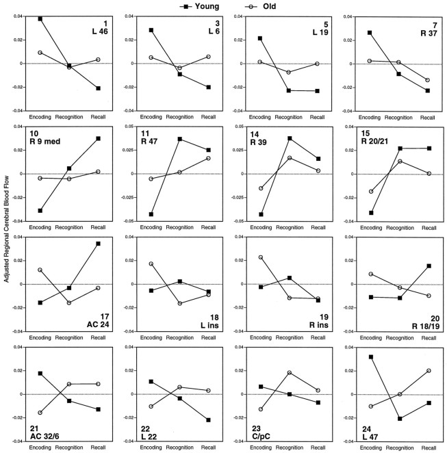
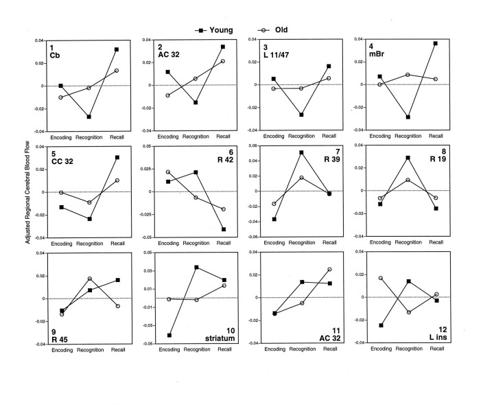
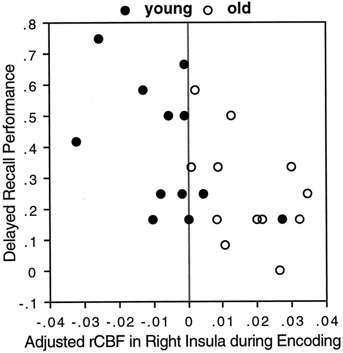
Positron emission tomography (PET) was used to compare regional cerebral blood flow (rCBF) in young (mean 26 years) and old (mean 70 years) subjects while they were encoding, recognizing, and recalling word pairs. A multivariate partial-least-squares (PLS) analysis of the data was used to identify age-related neural changes associated with (1) encoding versus retrieval and (2) recognition versus recall. Young subjects showed higher activation than old subjects (1) in left prefrontal and occipito-temporal regions during encoding and (2) in right prefrontal and parietal regions during retrieval. Old subjects showed relatively higher activation than young subjects in several regions, including insular regions during encoding, cuneus/precuneus regions during recognition, and left prefrontal regions during recall. Frontal activity in young subjects was left-lateralized during encoding and right-lateralized during recall [hemispheric encoding/retrieval asymmetry (HERA)], whereas old adults showed little frontal activity during encoding and a more bilateral pattern of frontal activation during retrieval. In young subjects, activation in recall was higher than that in recognition in cerebellar and cingulate regions, whereas recognition showed higher activity in right temporal and parietal regions. In old subjects, the differences in blood flow between recall and recognition were smaller in these regions, yet more pronounced in other regions. Taken together, the results indicate that advanced age is associated with neural changes in the brain systems underlying encoding, recognition, and recall. These changes take two forms: (1) age-related decreases in local regional activity, which may signal less efficient processing by the old, and (2) age-related increases in activity, which may signal functional compensation.
Figures
References
-
- Cabeza R, Nyberg L (1996) Imaging cognition: an empirical review of PET studies with normal subjects. J Cognit Neurosci, in press. - PubMed
-
- Cabeza R, Kapur S, Craik FIM, McIntosh AR, Houle S, Tulving E (1996) Functional neuroanatomy of recall and recognition: a PET study of episodic memory. J Cognit Neurosci, in press. - PubMed
Publication types
MeSH terms
LinkOut - more resources
Full Text Sources
Medical