. 1997 Jan 1;17(1):420-7.
doi: 10.1523/JNEUROSCI.17-01-00420.1997.

Nitric oxide production in rat thalamus changes with behavioral state, local depolarization, and brainstem stimulation

Affiliations

Nitric oxide production in rat thalamus changes with behavioral state, local depolarization, and brainstem stimulation

J A Williams et al. J Neurosci. .

Abstract

Since its discovery as a putative neurotransmitter in the CNS, several functional roles have been suggested for nitric oxide (NO). However, few studies have investigated the role of NO in natural physiology. Because NO synthase (NOS) has been localized in regions believed to be important for attention and arousal, we hypothesized that NO production would be state-dependent. To test this hypothesis, we used in vivo microdialysis, coupled with the hemoglobin-trapping technique, to monitor extracellular NO concentrations in rat thalamus during wake, slow-wave sleep (SWS), and rapid eye movement (REM) sleep. The thalamus is known to receive a massive innervation from the NOS/cholinergic neurons in the mesopontine brainstem, which have been suggested to play a key role in EEG desynchronized states. To test whether thalamic NO output was sensitive to neuronal-dependent changes in the mesopontine brainstem, we measured thalamic NO concentration in response to electrical stimulation in the laterodorsal tegmentum (LDT) of anesthetized rats. Finally, the calcium dependence of NO release was tested by local depolarization with a high potassium dialysate or by addition of a calcium chelator. The results showed that (1) extracellular NO concentrations in the thalamus were high during wake and REM sleep and significantly lower during SWS, (2) thalamic NO release increased in response to LDT stimulation in both a site-specific and tetrodotoxin (TTX)-dependent manner, and (3) NO production was calcium-dependent. These data suggest that thalamic NO production may play a role in arousal.


Figures

Fig. 1.
Fig. 1.
Calibration curve obtained by quantitative oxidation of increasing concentrations of HbO2 to metHb (0.5–5 μm) by aqueous NO. Values along they-axis are the absorbance differences between 577 and 591 nm obtained for metHb subtracted from that obtained from corresponding concentrations of HbO2. NO concentrations were estimated by using the slope of the line.
Fig. 2.
Fig. 2.
Histological verification of placement of microdialysis probes and stimulating electrodes. A, Composite photomicrograph of a cresyl violet-stained coronal section showing horizontal probe track. Arrows mark the position of the boundaries of the active surface of the membrane, which traversed the thalamus (see Materials and Methods). r, Reticular thalamic nucleus; v, ventrolateral complex;h, lateral habenula; p, posterior thalamic nucleus. Scale bar, 500 μm. B, NADPH-diaphorase-stained sagittal section showing the site of the stimulating electrode tip among the NOS-containing neurons in the laterodorsal tegmental nucleus. Rostral is left; dorsal is up. Scale bar, 100 μm.
Fig. 3.
Fig. 3.
On-line experiments demonstrating that the HbO2 oxidation measured in the samples was attributable to NO synthase (NOS) and Ca2+-dependent activity. In each panel, the mean ± SEM concentrations of extracellular NO in pmol/min are shown versus time in minutes. Dashed linesrepresent the baseline average. A, Intraperitoneal injection of the NOS inhibitor N-ARG significantly reduced baseline concentrations in awake animals (p < 0.001; n = 4).B, Application of a Ca2+ chelator BAPTA (10 mm) in a Ca2+-free dialysate solution significantly and reversibly reduced baseline concentrations in awake animals (p < 0.01; n = 3). C, Application of a solution containing 30 mm K+ significantly increased NO release in urethane-anesthetized rats (p < 0.001;n = 4). D, Addition of 10 mm BAPTA to the high K+ solution blocked the potassium-induced increase (n = 3), suggesting that the increase was attributable to Ca2+-dependent mechanisms. Although baselines varied slightly among groups, these differences were not significant between the animals within the awake groups (A, B) or anesthetized groups (C, D), but the baselines in anesthetized animals were significantly lower than those in awake animals (p < 0.001).
Fig. 4.
Fig. 4.
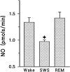
Extracellular NO concentrations across behavioral state in the rat thalamus. Mean NO concentrations (±SEM) are reported for wake (Wake), slow-wave sleep (SWS), and REM sleep (REM; n = 7). NO concentrations did not differ between wake and REM but were significantly lower during SWS. *p < 0.01.
Fig. 5.
Fig. 5.
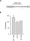
Effects of electrical stimulation of the laterodorsal tegmental nucleus (LDT) on cortical EEG and NO concentrations in the thalamus of anesthetized rats. A, EEG recording during a 10 sec stimulation pulse exhibits low amplitude waves (6–8 Hz), as compared with the high amplitude slow-wave activity during the interstimulus intervals (see Results).B, LDT stimulation significantly increased NO concentration (LDT Stim; n = 12; *p < 0.001). This effect was blocked by addition of 1 μm TTX to the Ringer’s solution (+TTX; n = 3). No change was observed when the stimulating electrode was placed in the cerebellum (Cb Stim; n = 3).

Similar articles

Cited by

References

    1. Abarca J, Gysling K, Roth RH, Bustos G. Changes in extracellular levels of glutamate and aspartate in rat substantia nigra induced by dopamine receptor ligands: in vivo microdialysis studies. Neurochem Res. 1995;20:159–169. - PubMed
    1. Adachi T, Inanami O, Sato A. Nitric oxide (NO) is involved in increased cerebral cortical blood flow following stimulation of the nucleus basalis of Meynert in anesthetized rats. Neurosci Lett. 1992;139:201–204. - PubMed
    1. Amir S, Robinson B, Edelstein K. Distribution of NADPH-diaphorase staining and light-induced fos expression in the rat suprachiasmatic nucleus region supports a role for nitric oxide in the circadian system. Neuroscience. 1995;69:545–555. - PubMed
    1. Bagetta G, Iannone M, Del Duca C, Nistico G. Inhibition by Nω-nitro-l-arginine methyl ester of the electrocortical arousal response in rats. Br J Pharmacol. 1993;108:858–860. - PMC - PubMed
    1. Balcioglu A, Maher TJ. Determination of kainic acid-induced release of nitric oxide using a novel hemoglobin trapping technique with microdialysis. J Neurochem. 1993;61:2311–2313. - PubMed

Publication types

LinkOut - more resources