. 1997 Jan 1;17(1):477-92.
doi: 10.1523/JNEUROSCI.17-01-00477.1997.

Changes in hippocampal circuitry after pilocarpine-induced seizures as revealed by opioid receptor distribution and activation

Affiliations

Changes in hippocampal circuitry after pilocarpine-induced seizures as revealed by opioid receptor distribution and activation

S B Bausch et al. J Neurosci. .

Abstract

The pilocarpine model of temporal lobe epilepsy was used to study the time-dependent changes in dentate gyrus circuitry after seizures. Seizures caused a decrease in mu- and delta-opioid receptor immunoreactive (MOR-IR and DOR-IR, respectively) neurons in the hilus and MOR-IR neurons in the granule cell layer. Additionally, diffuse DOR-IR, MOR-IR, and GABA immunoreactivities (GABA-IR) were increased in the inner molecular layer. Using the in vitro hippocampal slice preparation to study the physiological consequences of the anatomical changes, we found that the disinhibitory effects of the mu-opioid receptor agonist [D-Ala2, MePhe4,Gly-(ol)5]-enkephalin (DAMGO) and the GABAA receptor antagonist bicuculline were greatly depressed 5-13 d after pilocarpine injection but returned to control levels within 6 weeks. The amplitudes of monosynaptic evoked IPSCs and the effects of DAMGO on this parameter were also slightly decreased 5-13 d after pilocarpine injection but significantly increased at 6 weeks. DAMGO significantly decreased the mean amplitude of spontaneous IPSCs (sIPSCs) at 6 weeks after pilocarpine injection but not in controls. The delta-opioid receptor agonist [D-Pen2,5]-enkephalin (DPDPE) principally inhibited excitatory transmission in saline-treated animals without affecting either sIPSCs or evoked IPSCs. The DPDPE-induced inhibition of excitatory transmission became more pronounced at 6 weeks after pilocarpine injection. These results illustrate the anatomical reorganization and functional changes in dentate gyrus circuitry evident in an animal model of temporal lobe epilepsy and provide evidence of compensatory changes after trauma to the hippocampal formation.


Figures

Fig. 1.
Fig. 1.
Hilar cell loss and mossy fiber sprouting were seen after pilocarpine-induced seizures. Transverse sections from saline- (A, C) and pilocarpine- (B, D) treated rats were stained with either cresyl violet (A, B) or neo-Timm stain (C, D), as described in Materials and Methods. At 5 d to 6 weeks after injection, there was a marked decrease in the number of neurons in the hilus of pilocarpine (B) compared with saline-injected (A) rats. In the same animals, there was a pronounced increase in mossy fibers seen by neo-Timm staining in the inner molecular layer of animals injected with pilocarpine (D) compared with saline (C). All sections shown are from animals killed 6 weeks after injection. g, Granule cell layer;h, hilus; m, molecular layer. Scale bars (shown in A, C), 250 μm.
Fig. 2.
Fig. 2.
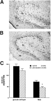
MOR-IR neurons were decreased in the hilus and granule cell layer of pilocarpine-treated compared with saline-treated rats. Transverse rat brain sections were stained with affinity-purified MT-2 (#2148) using the biotin amplification procedure, as described in Materials and Methods. Stained sections of the dentate gyrus show decreases in MOR-IR neurons in the hilus and the granule cell layer of pilocarpine- (B) compared with saline- (A) treated rats. Sections shown are from rats killed 19 d after injection. Compiled data illustrate decreases in (C) MOR-IR structures >10 × 10 μm and (D) MOR-IR somata in the hilus and granule cell layer of pilocarpine- compared with saline-treated rats. MOR-IR structures and MOR-IR somata localized in the subgranular region were included in the granule cell layer (see Materials and Methods). Data were compiled from 5 to 7 animals at each survival time in each treatment group. Data from different times were averaged, because there was no significant difference over time for either the saline or pilocarpine treatment group and no interaction between time and drug treatment. *Significant difference from saline-injected control rats (two- by three-way ANOVA, *p < 0.05; **p < 0.01).g, Granule cell layer; h, hilus;m, molecular layer. Scale bars (shown inA): A, B, 250 μm.
Fig. 3.
Fig. 3.
DOR-IR neurons were decreased in the hilus of pilocarpine- compared with saline-treated rats. Transverse rat brain sections were stained with affinity-purified DT-1 (#8663) using the biotin amplification procedure, as described in Materials and Methods. Stained sections of the dentate gyrus show a decrease in DOR-IR neurons in the hilus but not in the granule cell layer of pilocarpine- (B) compared with saline- (A) treated rats. Sections shown are from rats killed 14 d after injection. (C) Compiled data illustrate a decrease in DOR-IR structures in the hilus of pilocarpine- compared with saline-treated rats. Data were compiled from 6 to 9 animals at each survival time in each treatment group. Data from different survival times were averaged, because there was no significant difference over time for either the saline or pilocarpine treatment group and no interaction between time and drug treatment. *Significant difference from saline-injected controls (two- by three-way ANOVA, p < 0.05).g, Granule cell layer; h, hilus;m, molecular layer. Scale bars (shown inA): A, B, 250 μm.
Fig. 4.
Fig. 4.
Diffuse MOR-IR, GABA-IR, and DOR-IR were increased in the inner molecular layer of pilocarpine- compared with saline-treated rats. Transverse rat brain sections were stained with affinity-purified MT-2 (#2148) (A,B), anti-GABA antisera (C,D), or affinity-purified DT-1 (#8663) (E, F), using the biotin amplification procedure, as described in Materials and Methods. Immunoreactivity for all three antibodies was increased in the inner molecular layer of sections from pilocarpine- (B,D, F) compared with saline- (A, C, E) treated rats. Sections shown are from rats killed 8–9 d after injection. Sections from control and pilocarpine-treated animals were stained in parallel. All high-magnification photographs were taken from the upper blade near the open end of the granule cell layer. Arrowheads mark the edge of increased labeling. g, Granule cell layer;h, hilus; m, molecular layer. Scale bars (shown in A): AF, 100 μm.
Fig. 5.
Fig. 5.
Time-dependent changes were seen in the effects of the μ-opioid receptor agonist DAMGO and the GABAAreceptor antagonist bicuculline on dentate granule cell population spikes in pilocarpine-, compared with saline-treated, rats. Population spikes were recorded in normal ACSF, as described in Material and Methods. A, A representative stimulus response curve shows the increase in primary population spike amplitude caused by DAMGO (1 μm) or bicuculline (10 μm). (Rat killed 6 weeks after pilocarpine injection.) Inset shows placement of the stimulating (solid rectangle) and recording (open triangle) electrodes. B, Traces from a representative experiment illustrate the generation of a secondary population spike by DAMGO and bicuculline at a 300 μA stimulus. (Rat killed 14 d after saline injection.) Compiled data show time-dependent changes in the DAMGO-induced (1 μm) (C) and bicuculline-induced (10 μm) (E) increases in primary population spike amplitude evoked by a 300 μA stimulus, after pilocarpine-induced seizures. Compiled data show no changes in the DAMGO-induced (D) or bicuculline-induced (F) secondary population spike in the pilocarpine- compared with saline-injected rats. The amplitude of the secondary population spike was expressed as a percentage of the control primary population spike amplitude, both measured after a 300 μA stimulus. When a secondary population spike was observed in control recordings, only the drug-induced changes in the secondary population spike were included in the analysis. Opioid receptor-mediated effects were defined as the DAMGO effects that were reversible by 1 μm naloxone. Asterisk indicates a significant difference from saline-injected controls and 6 weeks after pilocarpine injection (one-way ANOVA, LSD post hoc comparison; p < 0.05).
Fig. 6.
Fig. 6.
Time-dependent changes were seen in the mean amplitude of monosynaptic evoked IPSCs and the DAMGO-induced depression of monosynaptic evoked IPSCs recorded in dentate granule cells after pilocarpine-induced seizures. Excitatory amino acid transmission was blocked with 50 μm APV and 10 μm CNQX. Putative postsynaptic opioid effects and the GABAB response were blocked with intracellular CsF and QX-314, as described in Materials and Methods. Granule cells were held at 0 mV for at least 30 sec before and after each monosynaptic IPSC data collection series.A, Traces from a representative experiment show that DAMGO (1 μm) decreased the amplitude of the monosynaptic IPSC evoked by both molecular layer (S1) and hilar (S2) stimulation measured in the same dentate granule cell. (Rat killed 6 weeks after saline injection.) Schematic shows placement of the bipolar stimulating (solid rectangles) and recording (open triangle) electrodes.B, C, Compiled data show time-dependent changes in the amplitude of the monosynaptic IPSC evoked from molecular layer (B) and hilar (C) stimulation in pilocarpine- compared with saline-treated rats. D,E, Compiled data show time-dependent changes in the DAMGO-induced (1 μm) depression of the monosynaptic IPSC evoked from molecular layer (D) and hilar (E) stimulation. Opioid receptor-mediated effects were defined as the DAMGO effects that were reversible by 1 μmnaloxone. Error bars indicate the mean ± SEM from the number of cells indicated in parentheses (11–19 animals per group) (B, C); or the number of animals indicated in parentheses (D,E). *Significant difference (one-way ANOVA, LSDpost hoc comparison; p < 0.05).
Fig. 7.
Fig. 7.
Effects of DAMGO on sIPSCs recorded in dentate granule cells were increased 6 weeks after pilocarpine-induced seizures. Recordings were done under the same conditions as described in Figure 6. Because recordings were done in the absence of the Na+ channel blocker tetrodotoxin (TTX), spontaneous events include both miniature IPSCs as well as action potential-dependent IPSCs. A, Traces from a representative experiment show that DAMGO (1 μm) decreased the frequency and amplitude of the sIPSCs. (Rat killed 6 weeks after pilocarpine injection.) Compiled data show no significant change in the mean sIPSC amplitude (B) or sIPSC frequency after pilocarpine-induced seizures (C). D, Compiled data show that DAMGO (1 μm) caused a greater depression of the mean sIPSC amplitude and sIPSC frequency 6 weeks after pilocarpine compared with saline injection. Opioid receptor-mediated effects were defined as the DAMGO effects that were reversible by 1 μmnaloxone. Error bars indicate the mean ± SEM from thenumber of cells indicated in parentheses(5–7 animals per group) (B,C) or thenumber of animals indicated inparentheses (D). *Significant difference from saline-injected controls (one-way ANOVA, LSD post hoccomparison; p < 0.05).
Fig. 8.
Fig. 8.
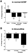
Time-dependent changes were seen in the effect of the δ-opioid receptor agonist DPDPE on the amplitudes of dentate granule cell population spikes after pilocarpine-induced seizures.A, Compiled data show time-dependent changes in primary population spike amplitude caused by DPDPE (1 μm) in normal ACSF in pilocarpine- compared with saline-injected rats. Population spikes were evoked by a 300 μA stimulus. B, Compiled data show seizure-induced, time-dependent changes in the effect of DPDPE (1 μm) on primary population spike amplitude in the presence of 10 μm bicuculline and high Mg2+ and high Ca2+ ACSF. Population spike amplitudes were measured at the control S1/2(stimulus intensity required to elicit a half-maximal response). Opioid receptor-mediated effects were defined as the DPDPE effects that were reversible by 1 μm naloxone or 100 nm −1 μm naltrindole. *Significant difference from saline-injected controls (Student’s t test,p < 0.05). **Significant difference from saline-injected controls and 5–13 d after pilocarpine injection (one-way ANOVA, LSD post hoc comparison;p < 0.01). #Significant effect based on a 99% confidence interval.
Fig. 9.
Fig. 9.
The δ-opioid receptor agonist DPDPE increased the amplitude of the monosynaptic IPSC recorded in dentate granule cells and evoked from hilar stimulation 6 weeks after pilocarpine injection. Recordings were done under the same conditions as described in Figure 6. A, B, Compiled data show no effect of DPDPE (1 μm) on the amplitude of the monosynaptic IPSC evoked by molecular layer stimulation (S1) (A). DPDPE (1 μm) did significantly increase the amplitude of the monosynaptic IPSC evoked by hilar stimulation (S2) (B) in rats killed 6 weeks after pilocarpine injection. Opioid receptor-mediated effects were defined as the changes in response caused by DPDPE that were reversible by 1 μm naloxone or 100 nm −1 μm naltrindole. *Significant difference from saline-injected controls (one-way ANOVA, LSD post hoccomparison; p < 0.05). #Significant effect based on a 99% confidence interval.

Similar articles

Cited by

References

    1. Adams JC. Biotin amplification of biotin and horseradish peroxidase signals in histochemical stains. J Histochem Cytochem. 1992;40:1457–1463. - PubMed
    1. Alreja M, Aghajanian GK. QX-314 blocks the potassium but not the sodium-dependent component of the opiate response in locus coeruleus neurons. Brain Res. 1994;639:320–324. - PubMed
    1. Amaral DG. A golgi study of cell types in the hilar region of the hippocampus in the rat. J Comp Neurol. 1978;182:851–914. - PubMed
    1. Andrade R. Blockade of neurotransmitter-activated K+ conductance by QX-314 in the rat hippocampus. Eur J Pharmacol. 1991;199:259–262. - PubMed
    1. Baez LA, Eskridge NK, Schein R. Postnatal development of dopaminergic and cholinergic catalepsy in the rat. Eur J Pharmacol. 1976;36:155–162. - PubMed

Publication types

MeSH terms