. 1997 Jan 15;17(2):607-14.
doi: 10.1523/JNEUROSCI.17-02-00607.1997.

Activation of hippocampal adenosine A3 receptors produces a desensitization of A1 receptor-mediated responses in rat hippocampus

Affiliations

Activation of hippocampal adenosine A3 receptors produces a desensitization of A1 receptor-mediated responses in rat hippocampus

T V Dunwiddie et al. J Neurosci. .

Abstract

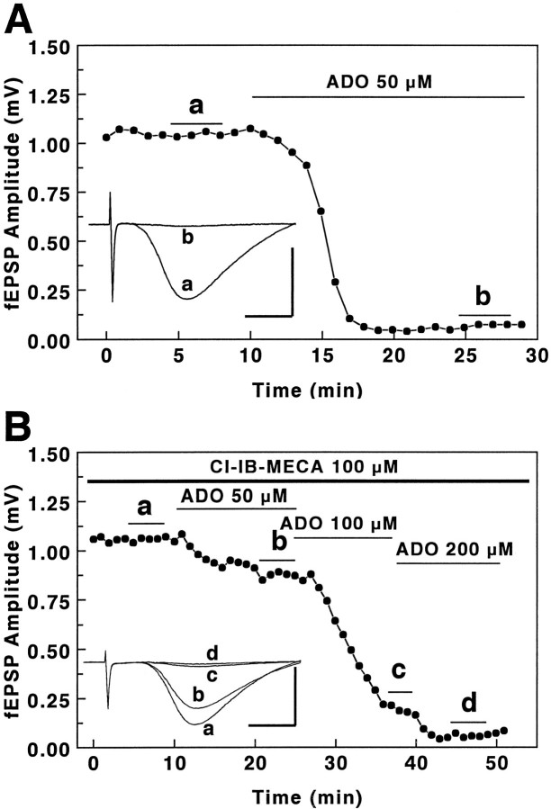
The adenosine A3 receptor is expressed in brain, but the consequences of activation of this receptor on electrophysiological activity are unknown. We have characterized the actions of a selective adenosine A3 receptor agonist, 2-chloro-N6-(3-lodobenzyl)-adenosine-5'-N-methyluronamide (Cl-IB-MECA), and a selective A3 receptor antagonist, 3-ethyl-5-benzyl-2-methyl-4-phenylethynyl-6-phenyl-1, 4-(+/-)-dihydropyridine-3,5-dicarboxylate (MRS 1191), in brain slices from rat hippocampus. In the CA1 region, activation of A3 receptors had no direct effects on synaptically evoked excitatory responses, long-term potentiation, or synaptic facilitation. However, activation of A3 receptors with Cl-IB-MECA antagonized the adenosine A1 receptor-mediated inhibition of excitatory neurotransmission. The effects of Cl-IB-MECA were blocked by pretreatment with MRS 1191, which by itself had no effect on A1 receptor-mediated responses. The presynaptic inhibitory effects of baclofen and carbachol, mediated via GABA(B) and muscarinic receptors, respectively, were unaffected by Cl-IB-MECA. The maximal response to adenosine was unchanged, suggesting that the primary effect of Cl-IB-MECA was to reduce the affinity of adenosine for the receptor rather than to uncouple it. Similar effects could be demonstrated after brief superfusion with high concentrations of adenosine itself. Under normal conditions, endogenous adenosine in brain is unlikely to affect the sensitivity of A1 receptors via this mechanism. However, when brain concentrations of adenosine are elevated (e.g., during hypoxia, ischemia, or seizures), activation of A3 receptors and subsequent heterologous desensitization of A1 receptors could occur, which might limit the cerebroprotective effects of adenosine under these conditions.


Figures

Fig. 1.
Fig. 1.
Effects of Cl-IB-MECA on hippocampal synaptic physiology. Slices were superfused with 100 nm(A) or 1 μm (B) Cl-IB-MECA, and the effects on PS (open circles) or fEPSP (filled circles) responses were determined. Results from individual slices are illustrated. Neither fEPSP nor PS responses were affected by treatment with either concentration of Cl-IB-MECA. On the other hand, superfusion with 30 μmadenosine (C) completely inhibited the PS and inhibited the fEPSP component of the response by ∼70%. On theright are signal averaged responses obtained before and during superfusion with Cl-IB-MECA (A, B) or adenosine (ADO, C). The top response of each pair is the control, the bottom in the presence of drug. PS responses recorded from the cell layer are shown above, and fEPSP responses from stratum radiatum are shownbelow. Adenosine eliminated the negative going PS response (C, top) and reduced the fEPSP (C, bottom).
Fig. 2.
Fig. 2.
Effects of Cl-IB-MECA on hippocampal synaptic plasticity. In A, slices were stimulated with a train of 100 Hz stimulation for 1 sec to induce LTP of the Schaffer collateral and commissural synapses. Under control conditions, this induced a reliable and persistent enhancement of the fEPSP amplitude. Pretreatment with 1 μm Cl-IB-MECA for 30 min before the stimulation train had no significant effect on the amplitude of the ensuing LTP. The ensemble averages for all the slices tested in this manner are illustrated in A. Billustrates hippocampal paired-pulse facilitation; when excitatory inputs to the CA1 region are stimulated twice in rapid succession, there is a significant potentiation of the second synaptic response called paired-pulse facilitation (Creager et al., 1980). Responses are illustrated from a control slice (B, a) and from a slice incubated in 1 μm Cl-IB-MECA (B, b) and tested with a 60 msec interpulse interval. The degree of facilitation (65 and 63%, respectively, in the examples shown) was not significantly different in the two conditions.
Fig. 3.
Fig. 3.
Cl-IB-MECA antagonizes the effects of adenosine on fEPSP responses. When slices were superfused repeatedly with 30 μm adenosine (A), the response to the second treatment with adenosine was an inhibition of the fEPSP amplitude comparable in magnitude to the first (i.e., there was no desensitization of the A1 receptor-mediated inhibition). However, when the slice was pretreated with either 100 nmor 1 μm Cl-IB-MECA before the second adenosine superfusion, the response was markedly inhibited (B). Similar effects were observed when the order was reversed, i.e., when the initial test was with Cl-IB-MECA + adenosine, and then the adenosine was tested alone after washout of Cl-IB-MECA (data not shown). Ensemble averages are shown in C for all three conditions. The inhibition of the adenosine response by 100 nm Cl-IB-MECA was statistically significant (p < 0.05), as was the inhibition by 1 μm Cl-IB-MECA (p < 0.0001).
Fig. 4.
Fig. 4.
MRS 1191 selectively blocks A3receptor mediated responses. A, Slices were superfused with adenosine, MRS 1191, and Cl-IB-MECA as denoted by thebars at the bottom of the figure, while the fEPSP response was tested at 15 sec intervals. Superfusion with 10 μm MRS 1191 had no effect on the adenosine response, but it blocked the ability of Cl-IB-MECA to disrupt adenosine responses (compare Fig. 3C). B, Summary of experiments with MRS 1191 and Cl-IB-MECA. Each bar represents the percent inhibition of the fEPSP response by 30 μmadenosine in the presence of the indicated drugs; slices were tested with the protocol illustrated in A or with a similar protocol but without MRS 1191. Each bar is the mean ± SEM for eight slices tested with an identical protocol. n.s., Not statistically significant, **p < 0.0001.
Fig. 5.
Fig. 5.
Cl-IB-MECA selectively disrupts the presynaptic modulatory effects of adenosine receptor agonists. Superfusion of slices with 30 μm adenosine, 20 nm NECA, 5 μm baclofen, or 5 μm carbachol (open bars) significantly inhibited the fEPSP response. Each bar shows the mean ± SEM inhibition of the response with each of the indicated drugs (the number of slices tested is shown to theleft of each pair of bars). Solid bars indicate slices that were pretreated for 30 min with 1 μm Cl-IB-MECA before superfusion with adenosine, NECA, baclofen, or carbachol; only the responses to adenosine and NECA were inhibited (**p < 0.001).
Fig. 6.
Fig. 6.
Effects of selective adenosine receptor agonists and antagonists on fEPSP responses. Slices were superfused with the indicated drugs alone (top three bars) or pretreated with the indicated drugs and then tested with 30 μmadenosine (cross-hatched bars). Each bar shows the mean ± SEM inhibition of the fEPSP response, and the number of slices tested is shown to the left of each bar. Neither the selective A3 agonist (Cl-IB-MECA) nor the A2a agonist (CGS 21680) had a significant effect on the fEPSP response. Pretreatment with 1 μm Cl-IB-MECA significantly attenuated the adenosine response, and this effect was not blocked by the selective A2a receptor antagonist CSC (1 μm). On the other hand, pretreatment with the A2a agonist CGS 21680 (100 nm) had no significant effect on the adenosine response.
Fig. 7.
Fig. 7.
Effect of Cl-IB-MECA on the adenosine dose–response curve. Mean dose–response curves are shown for adenosine alone, and adenosine + Cl-IB-MECA (1 μm). Eachpoint is the mean ± SEM response of at least five slices tested with the corresponding concentration of adenosine. EC50 values for the two conditions were 26 μmand 66 μm. The effect of Cl-IB-MECA on the EC50 value was statistically significant (p < 0.001), but the slopes of the corresponding dose–response curves (1.7, 1.6) were not significantly different. Note that the maximal effect of adenosine, which is normally a 95–100% inhibition of the fEPSP response, was not affected by Cl-IB-MECA.
Fig. 8.
Fig. 8.
A high concentration of Cl-IB-MECA reduces the potency of adenosine but not its maximal effect. Slices were incubated in a nonsuperfused slice chamber and were treated with adenosine alone (A) or pretreated with 100 μm Cl-IB-MECA and then tested with adenosine (B). Adenosine was added sequentially to achieve the indicated concentrations, but because the chamber was not superfused, washout was not possible with this experimental protocol. A illustrates an experiment with a control slice, which was pretreated with DMSO for 40 min before the beginning of the record (DMSO was used initially to dissolve the Cl-IB-MECA), and then adenosine was added; a concentration of 50 μm essentially eliminated the fEPSP response. InB, the slice was incubated in 100 μmCl-IB-MECA for 40 min (data not shown), then tested with increasing concentrations of adenosine. As in A, the fEPSP response could be completely inhibited, but the concentration of adenosine required was approximately fourfold higher in the presence of Cl-IB-MECA. The inset responses are signal averaged fEPSPs obtained during the periods indicated by the lettered bars. Calibration: 1 mV, 4 msec.
Fig. 9.
Fig. 9.
Desensitization induced by high concentrations of adenosine. A hippocampal slice was superfused initially with 20 μm adenosine, then the concentration was briefly increased to 1 mm, then reduced back to 20 μm, as indicated by the lines at thebottom. Signal averages of evoked fEPSPs in theinset correspond to responses evoked during the periods indicated by lettered line segments. The fact that the response recovered to a much higher level in 20 μmadenosine (d > b) and showed no change at all when the 20 μm adenosine superfusion was ended suggested that the inhibitory effects of this concentration of adenosine had been completely lost (a→b = 56% inhibition, vs e→d = 1% inhibition). The maximal response to adenosine did not appear to be affected during the 1 mm adenosine superfusion. The slight decrease in the baseline from a to e was not consistently observed.

References

    1. Abbracchio MP, Brambilla R, Ceruti S, Kim HO, von Lubitz DKJE, Jacobson KA, Cattabeni F. G protein-dependent activation of phospholipase C by adenosine A3 receptors in rat brain. Mol Pharmacol. 1995;48:1038–1045. - PubMed
    1. Ali H, Cunha-Melo JR, Saul WF, Beaven MA. Activation of phospholipase C via adenosine receptors provides synergistic signals for secretion in antigen-stimulated RBL-2H3 cells. Evidence for a novel adenosine receptor. J Biol Chem. 1990;265:745–753. - PubMed
    1. Creager R, Dunwiddie TV, Lynch GS. Paired-pulse and frequency facilitation in the CA1 region of the in vitro rat hippocampus. J Physiol (Lond) 1980;299:409–424. - PMC - PubMed
    1. Cunha RA, Constantino MD, Sebastião AM, Ribeiro JA. Modification of A1 and A2a adenosine receptor binding in aged striatum, hippocampus and cortex of the rat. NeuroReport. 1995;6:1583–1588. - PubMed
    1. Cunha RA, Johansson B, Constantino D, Sebastião AM, Fredholm BB. Evidence for high affinity binding sites for the adenosine A2A receptor agonist [3H]CGS21680 in the rat hippocampus and cerebral cortex that are different from striatal A2A receptors. Naunyn Schmiedebergs Arch Pharmacol. 1996;353:261–271. - PubMed

Publication types

MeSH terms

LinkOut - more resources