. 1997 Jan 15;17(2):615-24.
doi: 10.1523/JNEUROSCI.17-02-00615.1997.

Mechanisms of C5a and C3a complement fragment-induced [Ca2+]i signaling in mouse microglia

Affiliations

Mechanisms of C5a and C3a complement fragment-induced [Ca2+]i signaling in mouse microglia

T Möller et al. J Neurosci. .

Abstract

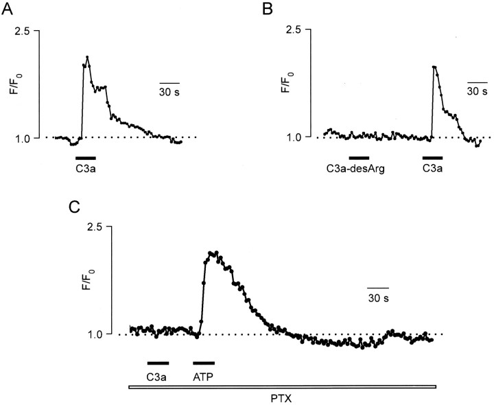
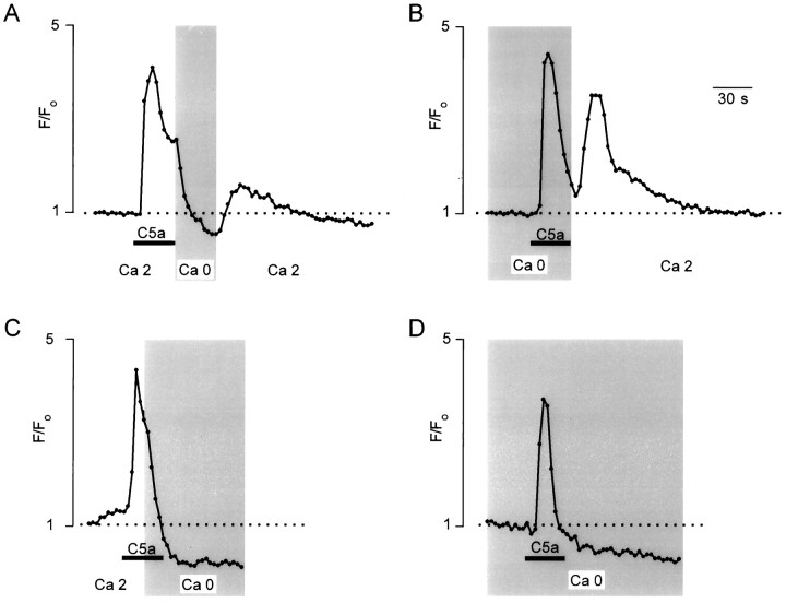
Microglial cells are activated in response to brain insults; the mechanisms of this process are not yet understood. One of the important signaling mechanisms that might be involved in microglia activation is related to changes in the intracellular calcium concentration ([Ca2+]i). Using fluo-3 microfluorimetry, we have found that external application of the complement fragment C5a (4-10 nM) induced [Ca2+]i elevation in microglial cells in situ in corpus callosum slices. Similarly, application of complement fragments C5a (0.1-10.0 nM) or C3a (100 nM) generates biphasic [Ca2+]i transients composed of an initial peak followed by a plateau in cultured microglia. Incubation of microglial cells for 30 min with pertussis toxin (PTX; 1 microgram/ml) inhibited both C5a- and C3a-triggered [Ca2+]i responses, suggesting the involvement of PTX-sensitive G-proteins in the signal transduction chain. Removal of Ca2+ ions from the extracellular solution eliminated the plateau phase and limited the response to the initial peak. The restoration of the extracellular Ca2+ concentration within 30-60 sec after the beginning of the complement fragment-induced [Ca2+]i elevation led to the recovery of the plateau phase. Inhibition of the endoplasmic reticulum Ca2+ pumps with 500 nM thapsigargin transiently increased the [Ca2+]i and blocked the [Ca2+]i signals in response to subsequent complement fragment application. Our data suggest that complement factors induce [Ca2+]i responses by Ca2+ release from internal pools and subsequent activation of Ca2+ entry controlled by the filling state of the intracellular Ca2+ depots.


Figures

Fig. 1.
Fig. 1.
C5a-induced [Ca2+]isignals in microglial cells in brain slices. A, Phase-contrast (left) and fluorescence image (right) taken from the same microglial cell visualized on the surface of a brain slice. Note that the cells sit on top of the slice surface. The focal plane was chosen to selectively record the fluorescence from the microglial cells. B, Representative examples of [Ca2+]i recordings from two different microglial cells in the brain slices in response to bath application of 5 nm C5a.
Fig. 2.
Fig. 2.
Representative examples of C5a-induced [Ca2+]i signals as recorded from different cultured microglial cells. A, Morphological appearance of cultured mouse microglial cells. B, Application of 2 nm C5a for 30 sec induced a biphasic [Ca2+]i transient, with an initial peak [Ca2+]i increase and a subsequent plateau phase of the [Ca2+]i elevation.C, Prolonged [Ca2+]i elevation triggered by 60 sec application of 2 nm C5a, which elicited a sustained plateau phase that did not recover to the resting level during the 5 min recording time. D, Blockade of the 2 nm C5a-induced [Ca2+]i signal by preincubation of cells with PTX (1 μg/ml, 30 min). Note that application of 100 μm ATP was still able to induce a [Ca2+]i elevation.
Fig. 3.
Fig. 3.
Concentration dependence of the C5a-mediated [Ca2+]i transients in cultured microglial cells. A, Examples of [Ca2+]itransients recorded from a microglial cell in response to application of 0.5 nm, 1 nm, 5 nm, and 10 nm C5a for 30 sec. B, Average values of the amplitudes of C5a-induced [Ca2+]i transients were obtained from nine experiments, similar to those described inA, and normalized to the amplitude of the response to 10 nm C5a. Error bars represent SD. C, Example of oscillatory [Ca2+]i response triggered by application of 0.5 nm C5a.
Fig. 4.
Fig. 4.
C3a-induced [Ca2+]isignals in cultured microglial cells. A, Application of 100 nm C3a for 30 sec induced [Ca2+]i elevation composed of an initial peak [Ca2+]i increase and a subsequent plateau phase of the [Ca2+]i elevation.B, Similar to A, [Ca2+]i was recorded from a single microglial cell in response to bath application of 100 nm C3a-desArg (nonactive analog of C3a) and 100 nm C3a. C, Blockade of the C3a-induced [Ca2+]i signal after preincubation of cells with PTX (1 μg/ml, 30 min). Control application of 100 μm ATP still induced [Ca2+]i elevation.
Fig. 5.
Fig. 5.
Complement fragments induced [Ca2+]i elevation in Ca2+-free external solution. A, C5a (2 nm)-induced [Ca2+]i transients were recorded in control conditions (left) and after removal of Ca2+ions from the bath (right). Note that in Ca2+-free extracellular solution, C5a was able to induce [Ca2+]i transients only once.B, The representative example of C3a (100 nm)-induced [Ca2+]i elevation as recorded in Ca2+-free external solution.
Fig. 6.
Fig. 6.
Modification of the C5a (2 nm)-induced [Ca2+]i signal by external Ca2+.A, Brief removal of the external Ca2+immediately after C5a application leads to an instant drop in [Ca2+]i; reestablishing the external Ca2+ caused the recovery of the sustained component of [Ca2+]i signal. B, Restoration of the physiological external Ca2+ concentration immediately after C5a application in Ca2+-free medium leads to the generation of the plateau phase. C, Persistent removal of the external Ca2+ after C5a application totally abolished the development of the plateau phase of [Ca2+]i signal. D, Application of C5a in Ca2+-free medium evoked only the peak component of the [Ca2+]i signal.
Fig. 7.
Fig. 7.
Thapsigargin inhibits [Ca2+]i mobilizing effects of C5a.A, In control conditions, external application of 2 nm C5a evoked a [Ca2+]i transient in a microglia cell. Subsequently, the cell was incubated with 500 nm thapsigargin. Application of thapsigargin induced [Ca2+]i elevation that returned to the basal level. The succeeding application of 2 nm C5a failed to induce the [Ca2+]i response.B, Similar to A, C5a and thapsigargin were applied while the cell was bathed in Ca2+-free extracellular solution. C, Effect of brief removal of extracellular Ca2+ on a thapsigargin (500 nm)-induced [Ca2+]i transient. Note that changing to the Ca2+-free extracellular media caused an immediate drop in the [Ca2+]ilevel, whereas restoration of the external Ca2+concentration to physiological levels resulted in recovery of the plateau phase of a thapsigargin-triggered [Ca2+]i transient.

References

    1. Bader M-F, Taupenot L, Ulrich G, Aumis D, Ciesielski-Treska J. Bacterial endotoxin induces [Ca2+]i transients and changes the organization of actin in microglia. Glia. 1995;11:336–344. - PubMed
    1. Barnum SR. Complement biosynthesis in the central nervous system. Crit Rev Oral Biol Med. 1995;6:132–146. - PubMed
    1. Berridge MJ. Inositol trisphosphate and calcium signalling. Nature. 1993;361:315–325. - PubMed
    1. Brockhaus J, Ilschner S, Banati RB, Kettenmann H. Membrane properties of ameboid microglial cells in the corpus callosum slice from early postnatal mice. J Neurosci. 1993;13:4412–4421. - PMC - PubMed
    1. Burger R, Bader A, Kirschfink M, Rotter U, Schrod L, Wörner I, Zilow G. Functional analysis and quantification of the complement C3 derived anaphylatoxin C3a with a monoclonal antibody. Clin Exp Immunol. 1987;68:703–709. - PMC - PubMed

Publication types

MeSH terms

LinkOut - more resources