. 1997 Jan 15;17(2):625-34.
doi: 10.1523/JNEUROSCI.17-02-00625.1997.

Bicuculline and gabazine are allosteric inhibitors of channel opening of the GABAA receptor

Affiliations

Bicuculline and gabazine are allosteric inhibitors of channel opening of the GABAA receptor

S Ueno et al. J Neurosci. .

Abstract

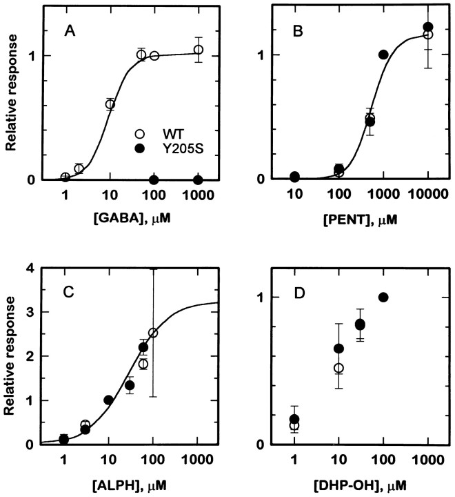
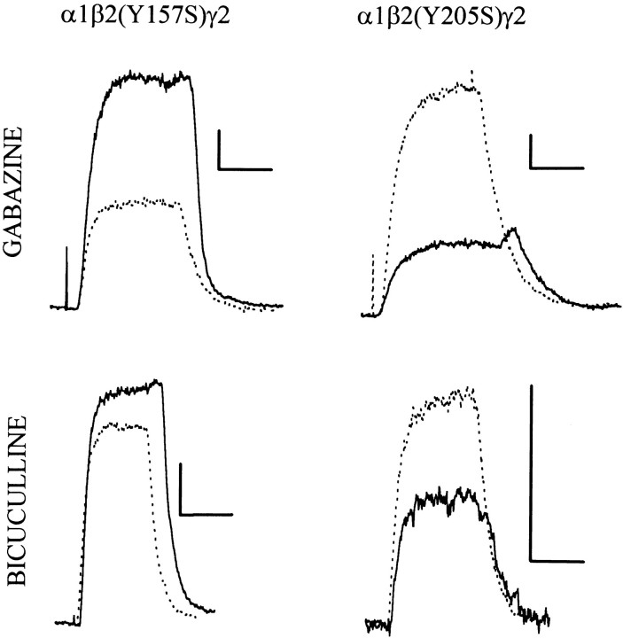
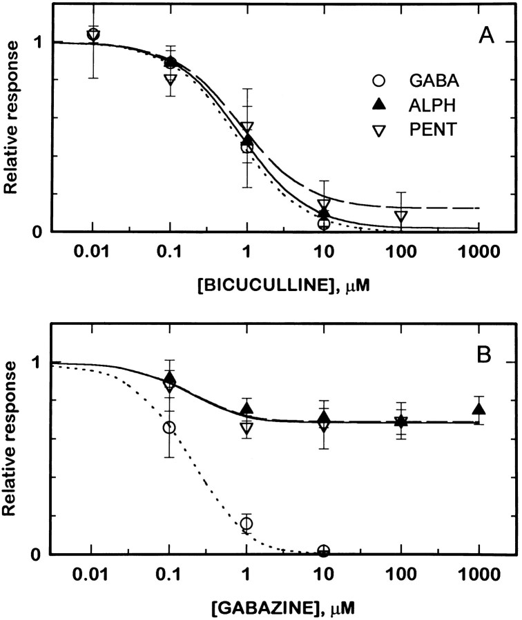
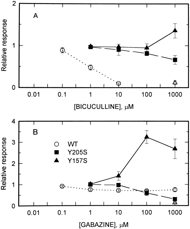
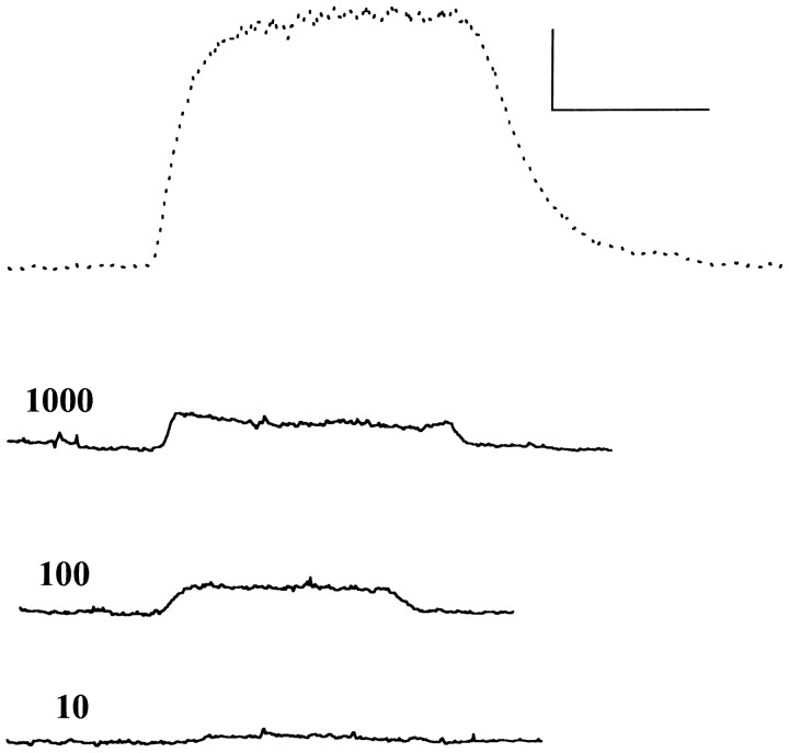
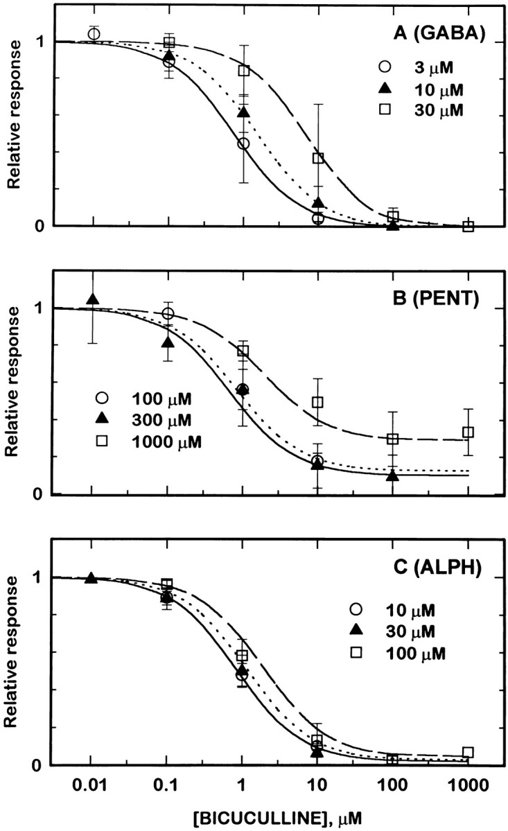
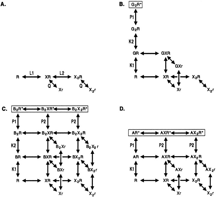
Anesthetic drugs are known to interact with GABAA receptors, both to potentiate the effects of low concentrations of GABA and to directly gate open the ion channel in the absence of GABA; however, the site(s) involved in direct gating by these drugs is not known. We have studied the ability of alphaxalone (an anesthetic steroid) and pentobarbital (an anesthetic barbiturate) to directly activate recombinant GABAA receptors containing the alpha 1, beta 2, and gamma 2L subunits. Steroid gating was not affected when either of two mutated beta 2 subunits [beta 2 (Y157S) and beta 2 (Y205S)] are incorporated into the receptors, although these subunits greatly reduce the affinity of GABA binding. These observations indicate that steroid binding and subsequent channel gating do not require these particular residues, as already shown for barbiturates. Bicuculline or gabazine (two competitive antagonists of GABA binding) reduced the currents elicited by alphaxalone and pentobarbital from wild-type GABAA receptors; however, gabazine produced only a partial block of response pentobarbital or alphaxalone, and bicuculline only partially blocked responses to pentobarbital. These observations indicate that the blockers do not compete with alphaxalone or pentobarbital for a single class of sites on the GABAA receptor. Finally, at receptors containing alpha 1 beta 2 (Y157S) gamma 2L subunits, both bicuculline and gabazine showed weak agonist activity and actually potentiated responses to alphaxalone. These observations indicate that the blocking drugs can produce allosteric changes in GABAA receptors, at least those containing this mutated beta 2 subunit. We conclude that the sites for binding steroids and barbiturates do not overlap with the GABA-binding site. Furthermore, neither gabazine nor bicuculline competes for binding at the steroid or barbiturate sites. The data are consistent with a model in which both gabazine and bicuculline act as allosteric inhibitors of channel opening for the GABAA receptor after binding to the GABA-binding site.


Figures

Fig. 1.
Fig. 1.
Activation of GABAA receptors containing β2 or β2(Y205S) subunits. Each panel shows concentration–response curves for an agonist: GABA (A), pentobarbital (PENT, B), alphaxalone (ALPH, C), and DHP-OH (D). In each panel, the open symbols show responses from QT6 cells transfected with α1β2γ2L subunits, whereas filled symbols show responses from cells transfected with α1β2(Y205S)γ2L subunits. GABA produced no gating of receptors containing the mutated subunit (points at 100 and 1000 μmGABA in A). For the other agonists tested, the data from receptors containing wild-type or mutated subunits were indistinguishable. The lines in A throughC show predictions derived from fitting an allosteric blocking model to data from receptors containing wild-type subunits (see Results). Symbols show mean for data from two to nine cells; error bars represent SD.
Fig. 4.
Fig. 4.
Responses to alphaxalone of cells expressing mutated β2 subunits. Each panel shows traces recorded from a cell exposed to 10 μm alphaxalone (dotted trace) and then to 10 μm alphaxalone plus 1 mm of an antagonist (solid trace). Actions of 1 mm gabazine are shown in the top rowand of 1 mm bicuculline in the bottom row. Cells transfected with α1β2(Y157S)γ2L subunits (left) showed potentiation between alphaxalone and either gabazine or bicuculline. Cells transfected with α1β2(Y205S)γ2L subunits showed block by either compound, but the block produced by bicuculline was reduced over that seen with wild-type receptors, whereas the block produced by gabazine was increased (see Figs. 3, 6). Calibration in each panel: 20 pA, 10 sec.
Fig. 2.
Fig. 2.
Action of blocking drugs on responses elicited from cells expressing α1β2γ2L receptors. Each panel shows traces recorded from a cell exposed to an agonist (dotted trace), or the same concentration of the agonist plus 10 μm of a blocking agent (solid trace). All cells were transfected with wild-type (α1β2γ2L) subunits. Theleft column shows the action of 10 μmbicuculline, whereas the right column shows the action of gabazine. Currents were elicited with 3 μm GABA (top row), 10 μm alphaxalone (middle row), or 300 μm pentobarbital (bottom row). Calibration in each panel: 20 pA, 10 sec. These records and those shown in Figures 4 and 5 were recorded at a holding potential of 0 mV, with a reversal potential for the responses of approximately −30 mV. Hence, the evoked currents are inward. Drugs were applied with a Y tube.
Fig. 3.
Fig. 3.
Bicuculline and gabazine block responses to GABA, pentobarbital, and alphaxalone. The agonists GABA (3 μm,open circles), alphaxalone (10 μm,filled triangles), and pentobarbital (300 μm, inverted open triangles) were applied to cells transfected with α1β2γ2L subunits, in the absence of a blocking drug and then in the presence of various concentrations of bicuculline (A) or gabazine (B). The figure shows the ratio of the response in the presence of a blocker to the response in the same cell in the absence of a blocker. The lines superimposed on the data (dotted lines, 3 μm GABA; solid lines, 10 μmalphaxalone; dashed lines, 300 μmpentobarbital) show predictions derived from fitting an allosteric blocking model to data from receptors containing wild-type subunits (see Results). Symbols show mean for data from two to six cells; error bars represent SD.
Fig. 6.
Fig. 6.
Actions of bicuculline and gabazine on alphaxalone-elicited responses of GABAA receptors containing mutated β2 subunits. Relative responses are shown to 10 μm alphaxalone applied to cells containing α1β2γ2L subunits (open circles, dotted lines), α1β2(Y205S)γ2L subunits (filled squares, dashed lines), or α1β2(Y157S)γ2L subunits (filled triangles, solid lines). The data obtained with bicuculline are shown in A, data with gabazine are shown inB. The lines simply connect the points. Also shown are the responses of receptors containing α1β2(Y157S)γ2L subunits to blocker applied in the absence of alphaxalone (open triangles). Symbols show mean for data from two to five cells; error bars represent SD .
Fig. 5.
Fig. 5.
Partial agonist action of gabazine on cells expressing α1β2(Y157S)γ2L subunits. The responses of a single cell to applications of 10 μm alphaxalone (top trace, dotted) and 1000, 100, and 10 μm gabazine alone. Calibration in the top panel: 20 pA and 10 sec for all traces.
Fig. 7.
Fig. 7.
The blocking effect of bicuculline depends on the concentration of agonist used. Cells transfected with wild-type receptors (α1β2γ2L subunits). Relative responses are shown for responses to three concentrations of agonist. For each concentration or agonist, the responses in the presence of a blocker are normalized to the response of that cell to the same concentration of agonist alone.A shows data obtained with GABA as agonist: 3 μm GABA (open circles, solid line), 10 μm GABA (solid triangles, dotted line), and 30 μm GABA (open squares, dashed line). B shows data obtained with pentobarbital as agonist: 100 μm pentobarbital (open circles, solid line), 300 μm pentobarbital (solid triangles, dotted line), and 1000 μmpentobarbital (open squares, dashed line).C shows data obtained with alphaxalone as agonist: 10 μm alphaxalone (open circles, solid line), 30 μm alphaxalone (solid triangles, dotted line), and 100 μm alphaxalone (open squares, dashed line). The lines superimposed on the data show predictions derived from fitting an allosteric blocking model to data from receptors containing wild-type subunits (see Results).
Fig. 8.
Fig. 8.
A diagram of the kinetic models used to assess the ability of a simplified allosteric model to describe the data. The models are described in Results. A shows the binding of blocker (X, bicuculline or gabazine) and the ensuing conformational change to the “dead” state (r). Note that the equilibrium constants (L1, L2, and Q) are omitted from B–D to simplify the figure. B–D show schemes for GABA, pentobarbital, and alphaxalone, respectively. Receptor states with open channels areboxed. B, GABA (G) was assumed to bind to the same two sites as blockers, so fewer heteroliganded forms of the receptor can occur. Furthermore, the channel can only activate when two GABA molecules are bound.C, Pentobarbital (B) was also assumed to bind to two sites, but in this case both pentobarbital and blockers can occupy sites on the same receptor. It was assumed that two pentobarbital molecules must be bound for a channel to activate and that dead receptors cannot activate. D, Alphaxalone (A) was assumed to bind to only a single site, but otherwise the scheme is identical to that for pentobarbital. Some binding steps (e.g., to the r state) are omitted for clarity in the figure.

Similar articles

Cited by

References

    1. Adams PR. Drug blockade of open end-plate channels. J Physiol (Lond) 1976;260:531–552. - PMC - PubMed
    1. Akaike N, Maruyama T, Tokutomi N. Kinetic properties of the pentobarbitone-gated chloride current in frog sensory neurones. J Physiol (Lond) 1987a;394:85–98. - PMC - PubMed
    1. Akaike N, Yakushiji T, Tokutomi N, Carpenter DO. Multiple mechanisms of antagonism of γ-aminobutyric acid (GABA) responses. Cell Mol Neurobiol. 1987b;7:97–103. - PMC - PubMed
    1. Amin J, Weiss DS. GABAA receptor needs two homologous domains of the β-subunit for activation by GABA but not by pentobarbital. Nature. 1993;366:565–569. - PubMed
    1. Arakawa O, Nakahiro M, Narahashi T. Chloride current induced by alcohols in rat dorsal root ganglion neurons. Brain Res. 1992;578:275–281. - PubMed

Publication types

MeSH terms

LinkOut - more resources