. 1997 Jan 15;17(2):667-75.
doi: 10.1523/JNEUROSCI.17-02-00667.1997.

Resetting the biological clock: mediation of nocturnal CREB phosphorylation via light, glutamate, and nitric oxide

Affiliations

Resetting the biological clock: mediation of nocturnal CREB phosphorylation via light, glutamate, and nitric oxide

J M Ding et al. J Neurosci. .

Abstract

Synchronization between the environmental lighting cycle and the biological clock in the suprachiasmatic nucleus (SCN) is correlated with phosphorylation of the Ca2+/cAMP response element binding protein (CREB) at the transcriptional activating site Ser133. Mechanisms mediating the formation of phospho-CREB (P-CREB) and their relation to clock resetting are unknown. To address these issues, we probed the signaling pathway between light and P-CREB. Nocturnal light rapidly and transiently induced P-CREB-like immunoreactivity (P-CREB-lir) in the rat SCN. Glutamate (Glu) or nitric oxide (NO) donor administration in vitro also induced P-CREB-lir in SCN neurons only during subjective night. Clock-controlled sensitivity to phase resetting by light. Glu, and NO is similarly restricted to subjective night. The effects of NMDA and nitric oxide synthase (NOS) antagonists on Glu-mediated induction of P-CREB-lir paralleled their inhibition of phase shifting. Significantly, among neurons in which P-CREB-lir was induced by light were NADPH-diaphorase-positive neurons of the SCN's retinorecipient area. Glu treatment increased the intensity of a 43 kDa band recognized by anti-P-CREB antibodies in subjective night but not day, whereas anti-alpha CREB-lir of this band remained constant between night and day. Inhibition of NOS during Glu stimulation diminished the anti-P-CREB-lir of this 43 kDa band. Together, these data couple nocturnal light, Glu, NMDA receptor activation and NO signaling to CREB phosphorylation in the transduction of brief environmental light stimulation of the retina into molecular changes in the SCN resulting in phase resetting of the biological clock.


Figures

Fig. 1.
Fig. 1.
Sensitivity of SCN to CREB phosphorylation by light or Glu is restricted to the night. Robust nuclear staining of P-CREB-lir was induced in the SCN under free-running conditions after stimulation with light in vivo (150 lux, 10 min) or Gluin vitro (10 mm in 0.2 μl drop, 10 min) during subjective night (CT 19–20). In contrast, only a weak basal level of P-CREB-lir was detected at CT 7 after the same light or Glu treatment. When the anti-P-CREB antibody was preabsorbed with equal amount of phospho-CREBtide, no positive immunoreactivity was detected. Scale bar, 200 μm.
Fig. 2.
Fig. 2.
Time course of P-CREB-lir in the SCN after light exposure. After a light pulse at CT 19, P-CREB-lir in the SCN reached maximum level within 10 min and remained at this level at 30 min. It declined to approximately half-maximum by 1 hr and returned to nearly basal level in ∼2 hr. In contrast, the P-CREB-lir in the SON of the same animals remained unchanged throughout the entire duration after the light exposure. General linear regression (GLM) for unbalanced ANOVA and post hoc test (Duncan) revealed that the levels of P-CREB-lir in the SCN at 10, 30, and 60 min groups are significantly different from basal level and from each other except for the values at 10 and 30 min. Each data point represents at least three animals; **p < 0.01.
Fig. 3.
Fig. 3.
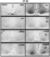
Blockade of Glu-induced P-CREB-lir in the SCN brain slices by inhibitors of the NMDA receptor and NOS. A 20 min preincubation in the NMDA receptor blocker APV (0.1 mm) or the NOS inhibitor, l-NAME (0.1 mm) diminished Glu-induced P-CREB-lir in the SCN at CT 20, whereas d-NAME (0.1 mm), the inactive stereoisomer of l-NAME, failed to block Glu-induced P-CREB-lir. These levels of staining are representative of four to six replications of each condition. Scale bar, 200 μm.
Fig. 4.
Fig. 4.
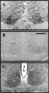
CT-dependent P-CREB induction by exogenous NO donor in the SCN. A microdrop (0.2 μl) of NO generator SNAP (0.01 mm) applied directly to the SCN induced P-CREB-lir at CT 19 (A), but not at CT 7 (B). Nissl stain demonstrates the histological boundary of the rat SCN (C). Scale bar, 200 μm.
Fig. 5.
Fig. 5.
Circadian sensitivity of SCN to phase resetting. Phase resetting was assessed by measuring the time-of-peak of the endogenous circadian rhythms of the neuronal activity of the SCN in brain slice. Top panel, Continuous single-unit extracellular recording of the unperturbed SCN neuronal activity from 112 units over 38 hr. The firing rate of this circadian rhythm peaked in midsubjective day at CT 7, on both days 2 and 3 in vitro. Middle panel, At CT 19, Glu advanced the peak of the SCN activity rhythm by 3 hr. A 0.2 μl droplet of 10 mm Glu was directly applied to the SCN for 10 min (arrow), followed by EBSS wash. Neuronal activity was recorded over 10–12 hr on each of the next two cycles to define the time-of-peak activity. Bottom panel, At CT 19, a 0.2 μl droplet of 0.01 mm SNAP advanced the peak of the SCN firing rate by 3.5 hr. The horizontal bars indicate the subjective night of the circadian cycle. The dashed vertical lines mark the time (CT 7) of the normal peak of the circadian rhythm of the neuronal activity in unperturbed and EBSS-treated controls.
Fig. 6.
Fig. 6.
Quantitative comparison of phase shifts and P-CREB-lir induced by various reagents affecting the Glu/NMDA receptor/NOS/NO pathway at CT 19–20. All measurements were made on SCN studied in vitro. Phase shifting data are replotted fromDing et al. (1994b) to facilitate direct comparison. P-CREB-lir was determined after treatment, fixation, and sectioning so thatn = each SCN brain slice per treatment (1 SCN slice was obtained from each animal). GLM for unbalanced ANOVA andpost hoc test (Duncan) revealed that only Glu, SNAP, and Glu + d-NAME significantly induced phase shifts as well as increased P-CREB-lir from basal levels in the SCN in vitro. Each data point represents the mean ± SD of four to eight animals; **p < 0.01.
Fig. 7.
Fig. 7.
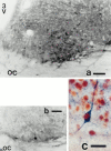
Colocalization of NADPH diaphorase and light-induced P-CREB-lir in the SCN. NADPH-diaphorase-positive neurons of varying size, arborization, and intensity are localized in the ventrolateral region of the SCN (a). Some diaphorase-containing neurons in the ventrolateral SCN have long processes projecting to the core of the SCN (b). The double-label immunocytochemistry and histochemistry revealed that the brown nuclear staining of P-CERB-lir was colocalized with the blue cytoplasmic staining of neuronal diaphorase within SCN neurons (c). Scale bars: a, 50 μm;b, 40 μm; c, 20 μm.
Fig. 8.
Fig. 8.
Glu induces a 43 kDa phosphoprotein in the SCN at night. Top panel, Affinity-purified anti-P-CREB antibody was used in Western blot analysis of the SCN nuclear extracts. A robust increase above basal level of a 43 kDa band was detected after Glu treatment at CT 20, but not at CT 7. A less intense reaction was also seen in bands of 33–36 kDa. The NOS inhibitor l-NAME diminished the amount of the 43 kDa band recognized by the anti-P-CREB antibody. Bottom panel, When the same gel was reprobed with the anti-αCREB antibody, equivalent amounts of the 43 kDa protein were present under all conditions. All experiments were repeated at least three times.

Similar articles

Cited by

References

    1. Alberini CM, Ghiradi M, Metz R, Kandel ER. C/EBP is an immediate-early gene required for the consolidation of long-term facilitation. Cell. 1994;76:1099–1114. - PubMed
    1. Amir S, Robinson B, Edelstein K. Distribution of NADPH-diaphorase staining and light-induced Fos expression in the rat suprachiasmatic nucleus region supports a role for nitric oxide in the circadian system. Neuroscience. 1995;69:545–555. - PubMed
    1. Bading H, Ginty DD, Greenberg ME. Regulation of gene expression in hippocampal neurons by a distinct calcium signaling pathway. Science. 1993;260:181–186. - PubMed
    1. Berkowitz LA, Riabowol KT, Gilman MZ. Multiple sequence ele-ments of a single functional class are required for cyclic AMP responsiveness of the mouse c-fos promoter. Mol Cell Biol. 1989;9:4272–4281. - PMC - PubMed
    1. Bourtchuladze R, Frenguelli B, Blendy J, Cioffi D, Schutz G, Silva AJ. Deficient long-term memory in mice with a targeted mutation of the cAMP-responsive element-binding protein. Cell. 1994;79:59–68. - PubMed

Publication types

MeSH terms

LinkOut - more resources