. 1997 Jan 15;17(2):796-803.
doi: 10.1523/JNEUROSCI.17-02-00796.1997.

Increased probability of GABA release during withdrawal from morphine

Affiliations

Increased probability of GABA release during withdrawal from morphine

A Bonci et al. J Neurosci. .

Abstract

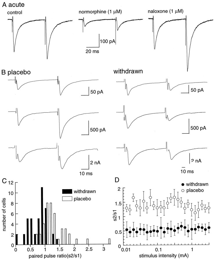
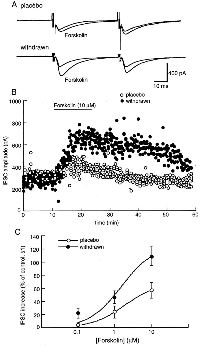
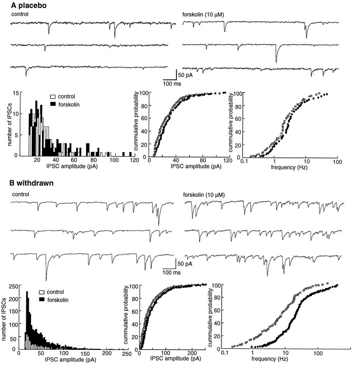
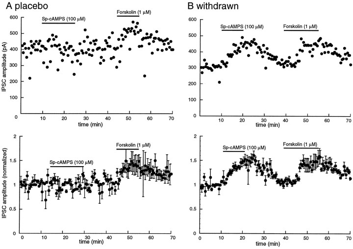
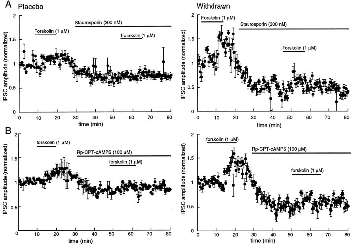
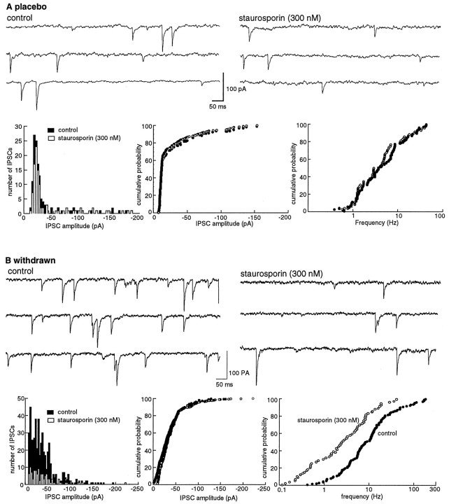
Opioid receptors located on interneurons in the ventral tegmental area (VTA) inhibit GABA(A)-mediated synaptic transmission to dopamine projection neurons. The resulting disinhibition of dopamine cells in the VTA is thought to play a pivotal role in drug abuse; however, little is known about how this GABAA synapse is affected after chronic morphine treatment. The regulation of GABA release during acute withdrawal from morphine was studied in slices from animals treated for 6-7 d with morphine. Slices containing the VTA were prepared and maintained in morphine-free solutions, and GABAA IPSCs were recorded from dopamine cells. The amplitude of evoked IPSCs and the frequency of spontaneous miniature IPSCs measured in slices from morphine-treated guinea pigs were greater than placebo-treated controls. In addition, activation of adenylyl cyclase, with forskolin, and cAMP-dependent protein kinase, with Sp-cAMPS, caused a larger increase in IPSCs in slices from morphine-treated animals. Conversely, the kinase inhibitors staurosporine and Rp-CPT-cAMPS decreased GABA IPSCs to a greater extent after drug treatment. The results indicate that the probability of GABA release was increased during withdrawal from chronic morphine treatment and that this effect resulted from an upregulation of the cAMP-dependent cascade. Increased transmitter release from opioid-sensitive synapses during acute withdrawal may be one adaptive mechanism that results from prolonged morphine treatment.


Figures

Fig. 1.
Fig. 1.
IPSCs from morphine-treated animals show paired-pulse depression, unlike those from controls, which show paired-pulse facilitation. A, Recording of GABAA-mediated IPSCs from a control slice using the paired-pulse protocol (left trace). Normorphine (1 μm) depressed the IPSC (middle trace), and that inhibition was reversed by the addition of naloxone to the normorphine-containing solution (right trace).B, Examples from three different cells in slices from both placebo- and morphine-treated animals. C, Cumulative results showing the distribution of paired-pulse ratio for many cells in slices from placebo (n = 41)- and morphine (n = 41)-withdrawn slices.D, The paired-pulse ratio is independent of the stimulus strength. These results are the average from four cells in each group of animals.
Fig. 2.
Fig. 2.
Forskolin augments the IPSC. This augmentation is significantly larger in morphine-withdrawn slices than in the placebo controls (p < 0.05). A, Examples of experiments from placebo- and morphine-treated animals.B, The amplitude of the first IPSC is plotted as a function of time in cells from placebo (open circles)- and morphine (solid circles)-withdrawn slices. Results are from four slices for each group. Forskolin (10 μm) increased the amplitude of the IPSC to a greater extent and for a more prolonged period in morphine-withdrawn slices than in controls.C, The concentration response to forskolin. The amplitude of the first IPSC in the paired-pulse protocol is plotted as a function of the concentration of forskolin. The amplitude was normalized against the mean of the first 10 IPSCs for each cell. Results were obtained from four cells from four different animals in each group. The data were fit with a least-squares regression from a logistic equation and gave estimates of the EC50 and maximum effect of forskolin of 1.8 μm and 66% increase in control and 1.5 μm and 125% increase in morphine-withdrawn slices.
Fig. 3.
Fig. 3.
Forskolin increased the frequency of spontaneous mIPSCs to a greater extent in morphine-withdrawn slices than in placebo controls. A, Experiment from a cell in a slice from a control animal. Top traces show the occurrence of spontaneous IPSCs in control (left) and after superfusion with forskolin (10 μm, right). The three plots below the traces show an amplitude histogram (left), a cumulative probability plot of the amplitude (middle), and a cumulative probability plot of the frequency of spontaneous IPSCs from the same cell shown above. In this particular cell, forskolin had little effect on the rate and amplitude of the spontaneous IPSCs. B, Illustration of the same experiment in a cell recorded in a slice taken from a morphine-treated animal. The initial rate of activity was higher, and forskolin induced a significant increase in the rate of spontaneous IPSCs (p < 0.05). All experiments were carried out in the presence of TTX (500 nm), CNQX (10 μm), APV (100 μm), strychnine (1 μm), and eticlopride (100 nm).
Fig. 4.
Fig. 4.
The cAMP analog Sp-cAMPS is more effective in increasing the IPSC in slices from morphine-withdrawn slices than placebo controls. The amplitude of the electrically evoked IPSC is plotted as a function of time. A, Top, Example of one experiment in a slice taken from a control animal. The cAMP analog Sp-cAMPS (100 μm) has no effect on the amplitude of the IPSC, whereas a low concentration of forskolin (1 μm) increased the amplitude of the IPSC.Bottom, Average of four such experiments in slices taken from four different animals. The average amplitude of IPSCs over the first 5 min was used to normalize the data. B,Top, An experiment in a single cell taken from a morphine-treated animal. In this case, cAMP analog Sp-cAMPS caused an increase in the IPSC that was about the same as that induced by forskolin. Bottom, Normalized and averaged results from four experiments.
Fig. 5.
Fig. 5.
The protein kinase inhibitors staurosporine (A, 300 nm) and Rp-CPT-cAMPS (B, 100 μm) decreased the IPSC in slices from both placebo- and morphine-treated animals. The inhibition is greater in morphine-withdrawn slices. In all plots, the IPSC amplitudes were normalized to the average determined over the first 5 min of the experiment. Plots on the left are from control animals (Placebo), and plots on the rightare from morphine-treated animals (Withdrawn).A, A low concentration of forskolin (1 μm) increased the amplitude of the IPSC, and staurosporine decreased the amplitude of the IPSC and blocked the forskolin-induced augmentation (n = 4). The plot labeled Withdrawnis the same experiment as shown at the left in slices taken from four morphine-treated animals. The inhibition induced by staurosporine is significantly larger than in controls (p < 0.05). B, The cAMP analog Rp-CPT-cAMPS, a cAMP-dependent kinase inhibitor, produced a greater inhibition of the amplitude of IPSCs in slices from both withdrawn slices than placebo controls (n = 4 for each group; p < 0.05). The same protocol was used as described for A.
Fig. 6.
Fig. 6.
The frequency of mIPSCs is decreased by staurosporine (300 nm). The decrease is greater in morphine-withdrawn slices than in controls. A, Experiment from a cell in a slice from a control animal. Top traces show the occurrence of spontaneous IPSCs in control (left) and after superfusion with staurosporine (300 nm, right). The three plots below the traces show an amplitude histogram (left), a cumulative probability plot of the amplitude (middle), and a cumulative probability plot of the frequency of spontaneous IPSCs from the same cell shown above. Staurosporine had little effect on the rate and amplitude of the spontaneous IPSCs.B, Illustration of the same experiment in a cell from a morphine-withdrawn slice. The initial rate of activity was higher, and staurosporine induced a significant decrease in the rate of spontaneous IPSCs (p < 0.05).

References

    1. Aghajanian GK. Tolerance of locus coeruleus neurons to morphine and suppression of withdrawal response by clonidine. Nature. 1978;276:186–187. - PubMed
    1. Akaoka H, Aston-Jones G. Opiate withdrawal-induced hyperactivity of locus coeruleus neurons is substantially mediated by augmented excitatory amino acid input. J Neurosci. 1991;11:3830–3839. - PMC - PubMed
    1. Andrade R, Vandermaelen CP, Aghajanian GK. Morphine tolerance and dependence in the locus coeruleus: single cell studies in brain slices. Eur J Pharmacol. 1983;91:161–165. - PubMed
    1. Bonci A, Williams JT. A common mechanism mediates long-term changes in synaptic transmission after chronic cocaine and morphine. Neuron. 1996;16:631–639. - PubMed
    1. Bozarth MA, Wise RA. Intracranial self-administration of morphine into the ventral tegmental area in rats. Life Sci. 1981;28:551–555. - PubMed

Publication types

MeSH terms

Substances

LinkOut - more resources